【細胞凋亡&細胞染色 】Abbkine促銷優惠活動DM&產品資料
【細胞增殖&毒理&衰老】| DNA Damege損傷/細胞Stress | 細胞凋亡&細胞染色 | 發炎反應ELISA分析 | 膽固醇代謝/代謝試驗 | 生化檢測試劑盒 | 抗體蛋白純化&其它 | Tag 標籤單多株抗體


TUNEL細胞凋亡檢測試劑盒(綠色螢光) | Abbkine TUNEL Apoptosis Detection Kit 貨號 KTA2010
![]() 凋亡細胞(apoptotic cells)最容易測量的特徵之一,是細胞核酸酶(cellular nucleases)破壞基因組DNA (genomic DNA)。末端脫氧核苷酸轉移酶dUTP缺口末端標記(Terminal deoxynucleotidyl transferase dUTP nick end labeling , TUNEL)是一種通過標記凋亡過程中產生的雙鏈DNA斷裂中的3′-羥基末端(3′-hydroxyl termini)來檢測DNA片段化的方法。 TUNEL測定法依賴於DNA中存在的缺口,該缺口可通過TdT進行鑑定,TdT是一種酶,可催化添加有螢光素標記的dUTP的添加。
凋亡細胞(apoptotic cells)最容易測量的特徵之一,是細胞核酸酶(cellular nucleases)破壞基因組DNA (genomic DNA)。末端脫氧核苷酸轉移酶dUTP缺口末端標記(Terminal deoxynucleotidyl transferase dUTP nick end labeling , TUNEL)是一種通過標記凋亡過程中產生的雙鏈DNA斷裂中的3′-羥基末端(3′-hydroxyl termini)來檢測DNA片段化的方法。 TUNEL測定法依賴於DNA中存在的缺口,該缺口可通過TdT進行鑑定,TdT是一種酶,可催化添加有螢光素標記的dUTP的添加。
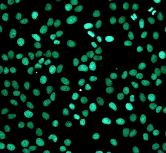
Fig. Fluorescence images using Abbkine TUNEL Apoptosis Detection Kit (Green Fluorescence) in HeLa cells with the treatment of 10 U/mL Dnase I for 10min.
產品描述: Abbkine TUNEL細胞凋亡檢測試劑盒(Abbkine TUNEL Apoptosis Detection Kit, 綠色螢光)依賴於DNA中存在的缺口,該缺口可通過TdT進行鑑定,TdT是一種酶,該酶催化添加螢光素標記的dUTP。該試劑盒為所有必需成分提供了優化的測定方案,適用於螢光酶標儀,螢光顯微鏡或流式細胞儀。在流行的FITC通道(Ex / Em = 490nm / 520 nm)處可以輕鬆檢測到其訊號。
- Kit components• TdT Enzyme • Equilibration Buffer (5X) • Label Nucleotide Mix
- Features & Benefits• Optimized assay protocol, suitable for fluorescence microplate reader, fluorescence microscope, or flow cytometer. • Signal can be easily detected at the popular FITC channel (Ex/Em = 490nm/520 nm).
- Usage notesThe reaction buffer contains cacodylate and cobalt chloride, highly toxic chemicals. After contact with skin, wash immediately with plenty of water. In case of accident or if you feel unwell, seek medical advice immediately. Do not drink, eat or smoke when using.
- Storage instructionsStable for at least 6 months at -20°C from date of shipment. Once opened, refer to list of materials supplied for storage conditions of individual components.
- ShippingGel pack with blue ice.
![]()
活細胞和死細胞雙染色套組 | CCK-8細胞增殖和細胞毒性試劑盒 | LDH細胞毒性測定試劑盒 | 細胞衰老檢測試劑盒(β-半乳糖苷酶 | 細胞增殖EdU Image試劑盒 | Abbkine 細胞膜染色 | 活細胞Lysosome染色
Annexin V-AbFluor™488細胞凋亡檢測試劑盒 | 貨號KTA0002
![]() 細胞凋亡(Apoptosis)是程序性細胞死亡(programmed cell death) 的一種形式,可以從組織中去除不需要的,受損的或衰老的細胞。在正常細胞中,負磷脂(phospholipids)駐留在細胞膜的內側,而膜的外表面則被不帶電荷的磷脂(uncharged phospholipids)(PS)佔據。細胞進入凋亡後,帶負電的PS從質膜(plasma membrane)的內部向外部leaflet轉運,從而使PS暴露於外部細胞環境。人類抗凝劑(human anticoagulant )-Annexin V是一種35-36 kDa的Ca2 +依賴性磷脂結合蛋白(Ca2+-dependent phospholipid-binding protein),對PS具有很高的親和力。用螢光團(fluorophore)或生物素(biotin)標記的膜聯蛋白V(Annexin V)可以通過與暴露在外部小葉上的PS結合來鑑定凋亡細胞(apoptotic cells)。碘化丙啶(PI)是一種不透過膜的DNA結合染料(membrane-impermeant DNA-binding dye),通常用於選擇性染色細胞群中的死細胞。PI被活細胞和早期凋亡細胞排除,但會對壞死和晚期凋亡細胞染色。 PI可以被488、532或546 n激發,並發出紅色螢光。
細胞凋亡(Apoptosis)是程序性細胞死亡(programmed cell death) 的一種形式,可以從組織中去除不需要的,受損的或衰老的細胞。在正常細胞中,負磷脂(phospholipids)駐留在細胞膜的內側,而膜的外表面則被不帶電荷的磷脂(uncharged phospholipids)(PS)佔據。細胞進入凋亡後,帶負電的PS從質膜(plasma membrane)的內部向外部leaflet轉運,從而使PS暴露於外部細胞環境。人類抗凝劑(human anticoagulant )-Annexin V是一種35-36 kDa的Ca2 +依賴性磷脂結合蛋白(Ca2+-dependent phospholipid-binding protein),對PS具有很高的親和力。用螢光團(fluorophore)或生物素(biotin)標記的膜聯蛋白V(Annexin V)可以通過與暴露在外部小葉上的PS結合來鑑定凋亡細胞(apoptotic cells)。碘化丙啶(PI)是一種不透過膜的DNA結合染料(membrane-impermeant DNA-binding dye),通常用於選擇性染色細胞群中的死細胞。PI被活細胞和早期凋亡細胞排除,但會對壞死和晚期凋亡細胞染色。 PI可以被488、532或546 n激發,並發出紅色螢光。
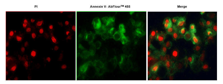
Fig.1. Hela cells were induced with camptothecin for 24 hours and stained with Annexin V- AbFluor™ 488 Apoptosis Detection Kit. The cell is a late stage apoptotic/necrotic cell with both Annexin V- AbFluor™ 488 and PI staining (green membrane with red fragmented nucleus).

Fig.2. Hela cells were induced with camptothecin for 24 hours and stained with Annexin V- AbFluor™ 488 Apoptosis Detection Kit. The combination of AbFluor™ 488 and propidium iodide allows for the distinction between early apoptotic cells (Annexin V- AbFluor™ 488 positive), late apoptotic and/or necrotic cells (Annexin V- AbFluor™ 488 and propidium iodide positive), and viable cells (unstained).
![]()
產品描述: Abbkine細胞凋亡檢測試劑盒 (Annexin V-AbFluor™488, 貨號KTA0002) 包含標有Abbkine專有綠色螢光染料的Annexin V-AbFluor™488,可通過流式細胞儀(flow cytometry)或螢光顯微鏡(fluorescence microscopy)對凋亡細胞(apoptotic cells)進行鑑定和定量。用AbFluor™488和碘化丙啶(propidium iodide , PI)對細胞進行同時染色可以區分完整細胞(intact cells),早期凋亡細胞(early apoptotic)和晚期凋亡細胞(late apoptotic)或壞死細胞(necrotic cells)。
| Product name | Annexin V-AbFluor™ 488 Apoptosis Detection kit (Green Fluorescence) |
| Applications notes | Abbkine Annexin V-AbFluor™ 488 Apoptosis Detection Kit contains Annexin V labeled with Abbkine proprietary green fluorescent dye AbFluor™ 488, which allows the identification and quantitation of apoptotic cells by flow cytometry or fluorescence microscopy. Simultaneous staining of cells with AbFluor™ 488 and propidium iodide (PI) allows the discrimination of intact cells, early apoptotic and late apoptotic or necrotic cells. |
| Alternative | Annexin 5; Annexin V; AbFluor™ 488 |
| Kit components | • Annexin V Binding Buffer (5x) • Annexin V- AbFluor™ 488 • Propidium Iodide (PI) |
| Features & Benefits | • Abbkine AbFluor™ 488 dye is superior to FITC as it is brighter, not affected by pH, and has much better photostability • Annexin V- AbFluor™ 488: Abs/Em = 491/517 nm; PI: Abs/Em = 535/617 nm (with DNA) • Suitable for flow cytometry or fluorescence microscopy |
| Storage instructions | Stable for at least 6 months at 4°C from date of shipment. Protect from light and do not freeze. |
| Shipping | Blue ice |
| Background | Apoptosis is a form of programmed cell death to remove unwanted, damaged, or senescent cells from tissues. In normal cells, the negative phospholipids reside on the inner side of the cellular membrane while the outer surface of the membrane is occupied by uncharged phospholipids (PS). After a cell has entered apoptosis, the negatively charged PS are transported from the inner to the outer leaflet of the plasma membrane, thus exposing PS to the external cellular environment. The human anticoagulant, Annexin V, is a 35–36 kDa Ca2+-dependent phospholipid-binding protein that has a high affinity for PS. Annexin V labeled with a fluorophore or biotin can identify apoptotic cells by binding to PS exposed on the outer leaflet. Propidium iodide (PI) is a membrane-impermeant DNA-binding dye that is commonly used to selectively stain dead cells in a cell population. PI is excluded by live cells and early apoptotic cells, but stains necrotic and late apoptotic cells with compromised membrane integrity. PI can be excited by the 488, 532, or 546 nm laser lines, and emits red fluorescence. |
JC-1線粒體膜電位測定試劑盒 | 貨號CAT # KTA4001
Mitochondrial Membrane Potential Assay Kit (JC-1)
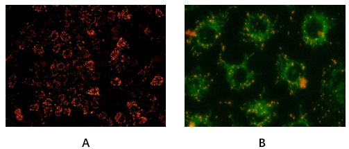
Fig. Hela cells stained with Abbkine Mitochondrial Membrane Potential Assay Kit (JC-1). A: Red fluorescence indicates healthy mitochondria, B: Green fluorescence indicates mitochondria in poor health (30min incubation in 20uM CCCP).
![]() Abbkine線粒體膜電位測定試劑盒(Mitochondrial Membrane Potential Assay Kit , JC-1貨號KTA4001)提供了一種簡單方法,基於碳菁染料(carbocyanine dye ) JC-1通過檢測線粒體跨膜電位的變化來區分健康細胞和凋亡細胞(apoptotic cells)。在健康細胞中,這種染料在粒腺體中積累(accumulates)和聚集(aggregates),形成鮮紅色的螢光團塊。耗散線粒體膜電位都會阻止JC-1染料在線粒體中積聚,因此該染料以其單體形式保留在細胞質中,導致從紅色的轉變(聚結的JC-1,Ex / Em = 585/590 nm)發出綠色螢光(JC-1單體,Ex / Em = 510/527 nm)。 JC-1可用作多種細胞類型(包括肌細胞和神經元)以及完整組織和分離的粒腺體中粒腺體潛力的指標。該試劑盒包含CCCP,CCCP導致質子梯度解偶聯,該質子梯度在電子傳輸鏈中電子載體的正常活動過程中建立,並因此消除了線粒體的電化學勢,可以用作防止JC-1聚集的對照組。
Abbkine線粒體膜電位測定試劑盒(Mitochondrial Membrane Potential Assay Kit , JC-1貨號KTA4001)提供了一種簡單方法,基於碳菁染料(carbocyanine dye ) JC-1通過檢測線粒體跨膜電位的變化來區分健康細胞和凋亡細胞(apoptotic cells)。在健康細胞中,這種染料在粒腺體中積累(accumulates)和聚集(aggregates),形成鮮紅色的螢光團塊。耗散線粒體膜電位都會阻止JC-1染料在線粒體中積聚,因此該染料以其單體形式保留在細胞質中,導致從紅色的轉變(聚結的JC-1,Ex / Em = 585/590 nm)發出綠色螢光(JC-1單體,Ex / Em = 510/527 nm)。 JC-1可用作多種細胞類型(包括肌細胞和神經元)以及完整組織和分離的粒腺體中粒腺體潛力的指標。該試劑盒包含CCCP,CCCP導致質子梯度解偶聯,該質子梯度在電子傳輸鏈中電子載體的正常活動過程中建立,並因此消除了線粒體的電化學勢,可以用作防止JC-1聚集的對照組。
![]()
| Product name | Mitochondrial Membrane Potential Assay Kit (JC-1) |
| Kit components | • JC-1 Stain • CCCP (10 mM) • Assay Buffer (5×) |
| Features & Benefits | • Measure mitochondrial membrane potential as an indicator of cell health. • Use JC-1, a fluorescent, lipophilic, cationic dye. • Red fluorescence indicates healthy mitochondria and green fluorescence indicates mitochondria in poor health. • Optimized for flow cytometery or fluorescent microscopy. • Contain CCC (Potent mitochondrial oxidative phosphorylation uncoupler). |
| Usage notes | • JC-1 Stain is difficult to prepare due to its low solubility. Make sure JC-1 Stain is completely thawed and warmed to room temperature before using. • JC-1 Stain is light senstive. Incubations need to be done in the dark. |
| Storage instructions | Refer to list of materials supplied for storage conditions of individual components. Stable for at least 12 months at recommended temperature from date of shipmen |
DAPI Staining Solution 粉狀 (DNA staining) | 貨號BMD00063
產品描述: DAPI是一種流行的藍色螢光DNA染料(blue fluorescent DNA dye)。該染料與dsDNA的結合,螢光增強約20倍。由於DAPI穿過完整的細胞膜,因此可用於染色活細胞和固定細胞。DAPI通常用於細胞凋亡檢測,染色後將使用螢光顯微鏡(fluorescence microscope)或流式細胞儀(flow cytometer)進行檢測。DAPI的藍色發射便於進行多重分析,因為DAPI與綠色螢光分子(如FITC和綠色螢光蛋白(GFP)或紅色螢光染料(如Texas Red)之間幾乎沒有螢光重疊。
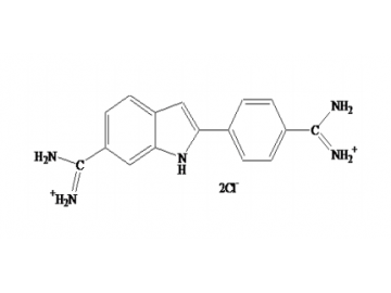
Fig. DAPI structure formula
![]()
| Product name | DAPI |
| CAS Number | 28718-90-3 |
| Molecular Formula | C16H17Cl2N5 |
| Alternative | DAPI,BMD00063 |
| Formulation | Yellow powder |
| Features & Benefits | • λEx/λEm: 360/460 nm (with DNA) |
| Molecular weight | 350.25 |
| Usage notes | • Soluble in H2O |
| Storage instructions | Store at -20°C and protect from light. Product is stable for at least 12 months from date of receipt if stored as recommended. |
| Shipping | Gel pack with blue ice. |
| Background | DAPI is a popular blue fluorescent DNA dye. The dye binds to the minor groove of dsDNA with approximately 20-fold fluorescence enhancement. DAPI is often applied to apoptosis detection, which will use fluorescence microscope or flow cytometer after staining. |
| Alternative | DAPI,BMD00063 |
引用文獻
Ruilian Li, Xianghua Yuan, Jinhua Wei, et al. Mar Drugs. 2019 Jan 10;17(1). pii: E43.
凋亡細胞分析(Annexin V法和TUNEL法) | Apoptosis Assay Cocktail貨號 KTD102-EN
凋亡細胞會產生許多重要的變化,包括細胞收縮(cell shrinkage)、細胞質凝集(cytoplasmic agglutination)、DNA斷裂(DNA disruption)和膜囊泡(membrane vesicles)的形成,並導致細胞成分分離形成凋亡小體(apoptotic dies),最終被巨噬細胞(macrophages)或實質細胞吞噬(parenchyma cells)Abbkine 基於新一代 AbFluor™ 螢光染色(AbFluor™ fluorescent staining)和高效重組蛋白技術(recombinant protein technology),開發了一種雞尾酒套裝,可同時檢測早期和晚期細胞凋亡的不同階段。 Vermes 是第一個使用 Annexin V 染色(Annexin V staining)特異性鑑定外翻細胞膜上的磷脂酰絲氨酸 (PS) 的研究人員,Annexin V 檢測被全球研究人員認為是最經典的方法。此外,DNA片段化(DNA fragmentation)是細胞凋亡晚期的一個重要特徵,基於DNA片段化檢測(DNA fragmentation detection)的TUNEL方法被認為是細胞凋亡研究的又一金標準。適用於組織(tissues)和細胞(cells)等不同樣品。高靈敏度(High sensitivity)、通用的細胞凋亡檢測試劑盒(universal apoptosis detection kit),可有效區分細胞凋亡(apoptosis)與壞死(necrosis)。同時,針對細胞凋亡檢測和結果的非標準化,我們開發了一種專利的細胞凋亡檢測陽性對照物質(positive control),並結合到細胞凋亡檢測雞尾酒中。本產品可作為細胞凋亡研究的標準化產品,能滿足大部分細胞凋亡檢測(apoptosis detection)的需要。是細胞凋亡檢測(apoptosis detection)的最佳選擇。 Abbkine開發了一種高靈敏度、通用的細胞凋亡試劑盒(universal apoptosis kit),可同時檢測早期和晚期細胞凋亡(apoptosis),適用於組織和細胞,有效區分細胞凋亡和壞死。本產品可作為細胞凋亡(apoptosis)研究的標準化產品,能滿足大部分細胞凋亡檢測的需要。是細胞凋亡檢測(apoptosis detection)的最佳選擇。

1 用於細胞凋亡研究的標準化試劑盒。
Annexin V法和TUNEL法作為細胞凋亡檢測的兩大金標準,幾乎涵蓋了細胞凋亡檢測的大部分要求。獨特的 Apoptosis Assay Cocktail 不僅包含上述兩種檢測方法,而且還擁有專利的細胞凋亡陽性對照,完美解決了細胞凋亡檢測和結果不規範的問題。
2 高靈敏度、通用的細胞凋亡檢測試劑盒。
優化後,該試劑盒可同時檢測早期和晚期不同階段的細胞凋亡,適用於組織、細胞等不同樣本,有效區分細胞凋亡和壞死。
3 廣泛使用。
可用於流式細胞儀和螢光顯微鏡觀察。

| Component name | 规格 |
| Annexin V-AbFluor™ 488 Apoptosis Detection Kit | 40 T |
| TUNEL Apoptosis Detection Kit (Green Fluorescence) | 20 T |
| Apoptosis Positive Control | 5 T |
HeLa cells were induced by inducers A and B as positive control group, and normal cells as negative control. Apoptosis detection cocktail kit was used for detection. Annexin V-Abfluor ™ 488 has a high affinity for PS, and PS exposed through the outer side of the cell binds to the membrane of cells in the early apoptotic stage, which were marked fluorescent green and the nucleus were marked red fluorescent by PI after structural damage.
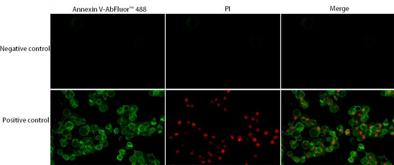
Fig.1 Annexin V-AbFluor™ 488 Apoptosis Detection Kit was used to detect the apoptosis effect of HeLa cells after apoptosis induction
After Annexin V-Abfluor ™ 488 labeling, the necrotic cells and apoptotic cells in early stage and late stage were accurately identified.
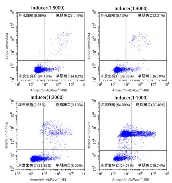
Fig.2 Flow cytometry was used to detect the apoptosis rate by different concentrations of inducer A and B.
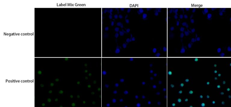
In the detection of apoptosis by TUNEL assay, deoxyribonucleotide terminal transferase (TdT) was used to label the deoxyribonucleotide – luciferase conjugates to the 3′- terminus of the DNA gap, and apoptosis was visualized.
Fig.3 TUNEL Apoptosis Detection Kit (Green Fluorescence) was used to detect the apoptosis effect of HeLa cells after apoptosis induction.
TraKine™Pro活細胞微管蛋白染色 | 貨號KTC4100
微管蛋白(Tubulin)是微管(microtubules.)的主要構建基塊。這種胞內圓柱形絲狀結構(cylindrical filamentous structure)幾乎存在於所有真核細胞(eukaryotic cells.)中。微管(Microtubules)在有絲分裂(mitosis),細胞內運輸(intracellular transport),鞭毛運動(flagellar movement)和細胞骨架(cytoskeleton.)中充當結構和移動元件。微管蛋白(Tubulin)是異二聚體(heterodimer),由α-微管蛋白(a-tubulin)和b-微管蛋白(b-tubulin)組成。兩個亞基的分子量均為55 kDa,並具有相當的同源性(homology)。研究最多的微管蛋白(tubulins)已從脊椎動物腦(vertebrate brains)中分離出來。可以在免疫螢光顯微鏡下觀察微管(microtubules),這使得能夠觀察超分子結構形式的蛋白質的細胞內組織。Tubulin is the major building block of microtubules. This intracellular cylindrical filamentous structure is present in almost all eukaryotic cells. Microtubules function as structural and mobile elements in mitosis, intracellular transport, flagellar movement, and in the cytoskeleton. Tubulin is a heterodimer, which consists of a-tubulin and b-tubulin; both subunits have a molecular weight of 55 kDa and share considerable homology. The most studied tubulins have been isolated from vertebrate brains. The microtubules can be viewed in immunofluorescent microscopy, which enables the observation of the intracellular organization of proteins that are in the form of a supramolecular structure.
產品描述:
TraKine™Pro是一系列長效型細胞影像染劑,可標記活細胞或固定之細胞的細胞內結構。 TraKine™Pro專有的優異螢光染料,其螢光範圍由UV可、見光至近IR光譜。另提供基於TraKine™Pro技術的客製化產品。 TraKine™Pro活細胞微管蛋白染色試劑盒(TraKine™ Pro Live-cell Tubulin Staining kit, KTC4100)(超高分辨率的綠色螢光, Ex / Em = 500/520 nm)是一種螢光成像工具,用於對哺乳動物活細胞中微管蛋白進行高特異性和低背景染色。探針為不透膜,但與buffer T incubation後,它可以通過囊泡區室(vesicular compartments)的內吞途徑(endocytosis pathway)進入細胞,隨後釋放到細胞質(cytoplasm)中。它特別適合於長期超分辨率成像。
![]()
- Kit components• TubGreen™ (200 uM)
• Buffer T - Features & Benefits• Optimized staining protocol for labeling Tubulin in mammalian living cells.
• Especially suitable for Confocal and long-term super-resolution imaging (such as SIM, STED, TIRF, STORM and PALM).
• Proprietary TubGreen™ (Ex/Em = 500/520 nm)-high specificity, low background and excellent photostability.
• Low levels of cytotoxicity. - Usage notesMake sure the pipette tips and PCR tubes were sterilized at high temperature and pressure. Make sure sterile environment and protect from light during the whole experiment.
- Storage instructionsRefer to list of materials supplied for storage conditions of individual components. Stable for at least 6 months at recommended temperature from date of shipment.
- ShippingGel pack with blue ice.
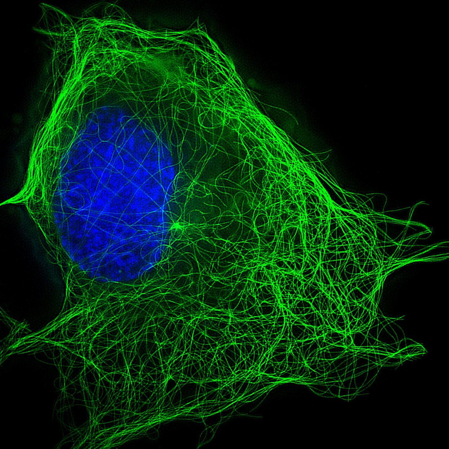
Fig.1. SIM imaging results of U2OS cells using Abbkine TraKine™ Pro Live-cell Tubulin Staining kit (Green Fluorescence with Super Resolution). It can be seen that the kit can specifically label microtubule structures in living cells and perform ultra-high resolution imaging.
| Product name | TraKine™ Pro Live-cell Tubulin-traker kit (Green Fluorescence) |
| Applications notes | TraKine™ Pro is series of long-term super-resolution cell staining imaging portfolio for labeling subcellular structures of live and fixed cells. TraKine™ Pro proprietary excellent fluorescent dyes span the full UV-visible and near IR spectrum. Customized products based on TraKine™ Pro technology are also available. TraKine™ Pro Live-cell Tubulin Staining kit (Green Fluorescence with Super Resolution) is a fluorescence imaging tool for staining of Tubulin in mammalian living cells with high specificity and low background. The proprietary probe in the kit consists of a microtubule recognition unit and a green fluorescent dye (Ex/Em=500/520 nm), the recognition unit can selectively recognize microtubules and binding with it. The whole probe is membrane impermeable, but after incubating with buffer T, it can enter cells through endocytosis pathway in vesicular compartments, and release into cytoplasm subsequently. It is especially suitable for long-term super-resolution imaging. |
| Kit components | •Tubulin Green (200 μM) •Buffer A (200 μM) •Buffer B (1 mM) |
| Features & Benefits | • Optimized staining protocol for labeling Tubulin in mammalian living cells. The product has been tested in U-2 OS,Hela,MCF-7 and MDA-MB-231 cell lines. U2OS cell line is preferred. If the sample type is not included in the above cell lines, we can provide experimental services for specific cell lines. • Suitable for Confocal high-definition imaging. • Suitable for structured light micro imaging (SIM) live cell research, ultra-high resolution microscopic imaging of live cells, and dynamic observation of cells in three-dimensional space. • Proprietary TubGreen™ (Ex/Em = 500/520 nm)-high specificity, low background and excellent photostability. • Low levels of cytotoxicity. |
| Usage notes | Make sure the pipette tips and PCR tubes were sterilized at high temperature and pressure. Make sure sterile environment and protect from light during the whole experiment. |
| Storage instructions | Refer to list of materials supplied for storage conditions of individual components. Stable for at least 6 months at recommended temperature from date of shipment. |
| Shipping | Gel pack with blue ice. |
| Precautions | The product listed herein is for research use only and is not intended for use in human or clinical diagnosis. Suggested applications of our products are not recommendations to use our products in violation of any patent or as a license. We cannot be responsible for patent infringements or other violations that may occur with the use of this product. |
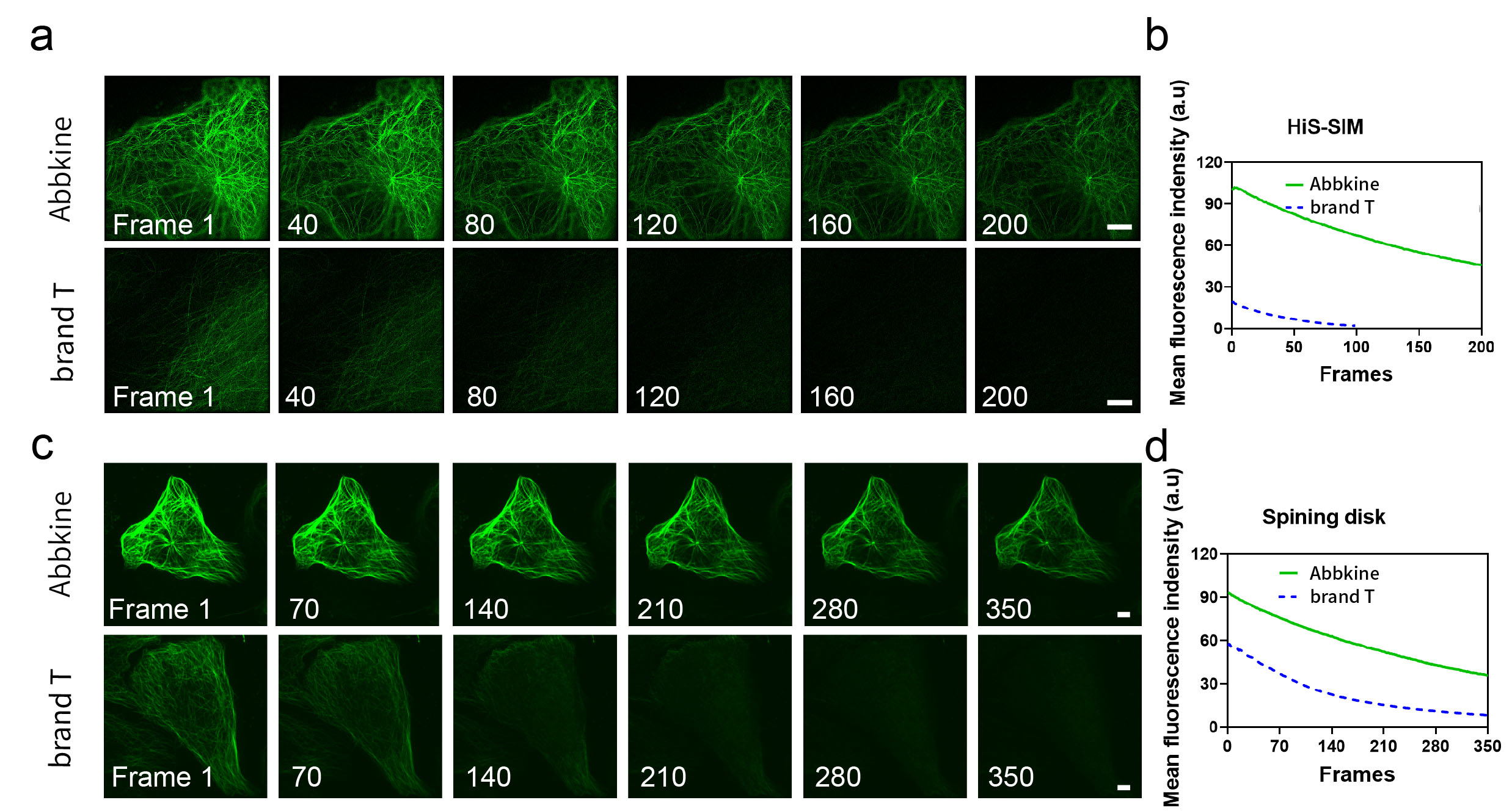
Fig.2. SIM imaging results of U2OS cells using Abbkine TraKine™ Pro Live-cell Tubulin Staining kit (Green Fluorescence with Super Resolution). It can be seen that the kit can specifically label microtubule structures in living cells and perform ultra-high resolution imaging.
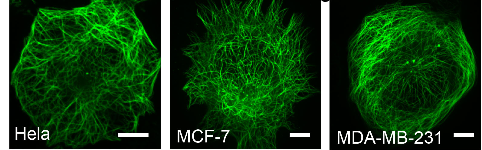
Fig.3. Confocal imaging results of U2OS cells using Abbkine TraKine™ Pro Live-cell Tubulin Staining kit (Green Fluorescence with Super Resolution). It shows that the kit has high labeling efficiency.
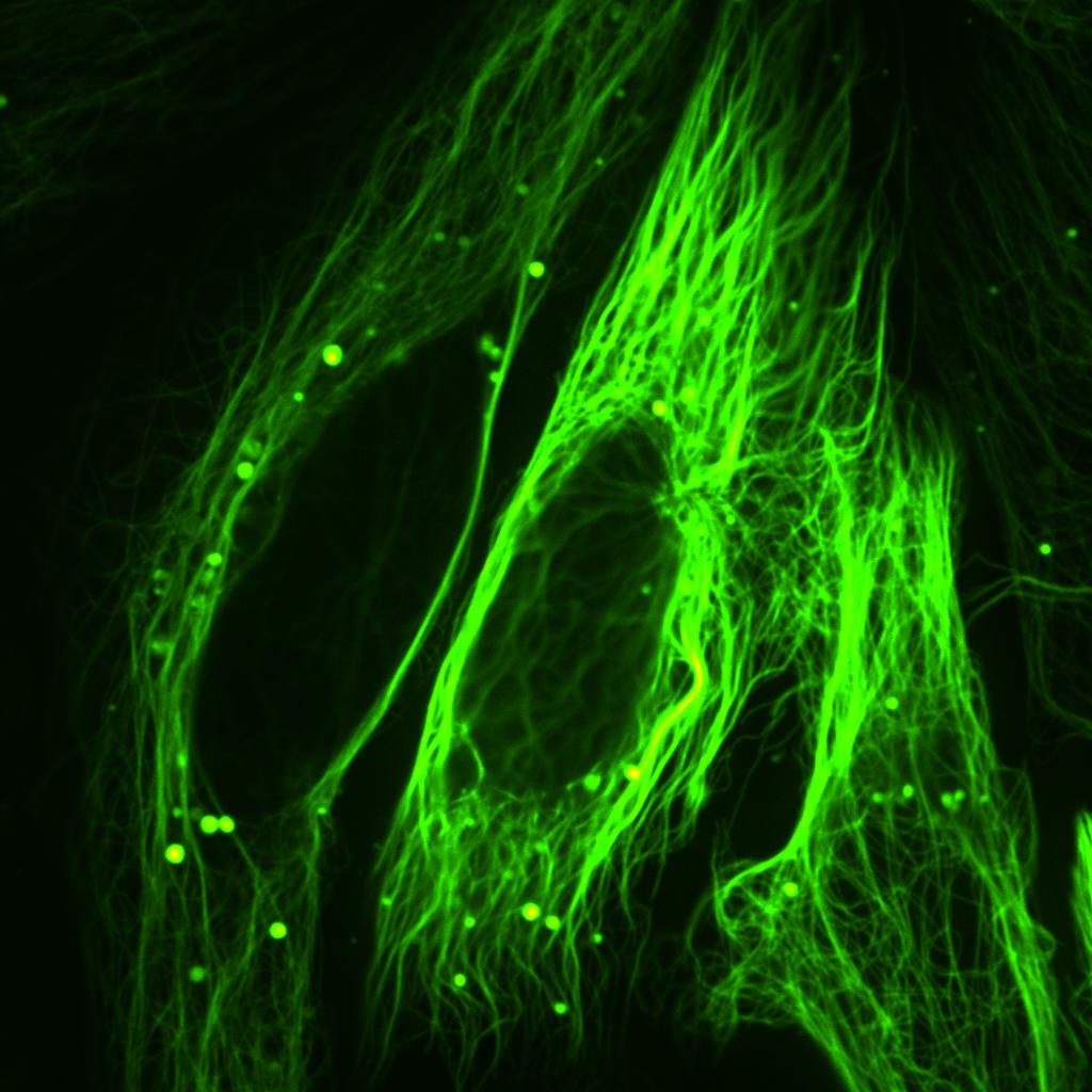
Fig.4. Confocal imaging results of U2OS cells using Abbkine TraKine™ Pro Live-cell Tubulin Staining kit (Green Fluorescence with Super Resolution). It shows that the kit has high labeling efficiency.
引用文獻
L Zhao, Y Jia, C Zhao. Acta biomaterialia, 2019 – Elsevier
Shuangshuang Wang, Jixuan Song, Yuchao Li, et al. Reactive and Functional Polymers. July 2019, 140: 48-55.
Abbkine 細胞膜染色 | Cell Plasma Membrane Staining Kits貨號KTC4001
細胞膜(plasma membrane)是一種薄的半滲透膜(semi-permeable membrane) ,由帶有嵌入蛋白質的脂質雙層(lipid bilayer)層組成,將所有細胞的內部與環境分離開來。細胞膜的基本功能是保護細胞免受周圍環境的影響。細胞膜控制物質進出細胞和細胞器的運動。通過這種方式,它被選擇性地滲透到離子和有機分子中。細胞膜參與各種細胞過程,如細胞粘附(cell adhesion)、電電導率(ion conductivity)和細胞信號(cell signaling),並作為多個細胞外結構的附著表面(attachment surface),包括細胞壁(cell wall)、glycocalyx和細胞內細胞骨架(intracellular cytoskeleton)。
Fig. Hela cells stained with TraKine™ Cell Plasma Membrane Staining Kit (Green Fluorescence).
產品描述: Abbkine 細胞膜染色試劑盒(Cell Plasma Membrane Staining Kits, KTC4001)是一套螢光成像工具(fluorescence imaging tools),可應用於活細胞、固定懸浮、貼附細胞,根據細胞類型和實驗條件,快速進行膜蛋白染色。該試劑盒使用專有的親脂性銀骨黃素染料(lipophilic carbocyanine dye , Ex/Em = 484/501 nm),在水中螢光微弱, 融入膜後具高度螢光及穩定性。
![]()
- Kit components
- CPM™ Green (1000×)
• Assay Buffer (10×) - Features & Benefits
- Enables the uniform staining of cell membrane across a wide variety of mammalian cell types, and can be used for both living and fixed suspended or attached cells.
• Optimized for various fluorescence platforms such as microplate assays, flow cytometry and fluorescence microscope.
• Proprietary CPM Green™ (Ex/Em = 484/501 nm)-resulting in accelerated diffusion within membranes, staining is also maintained after fixation with formaldehyde, enabling further multi-color staining. - Storage instructions
Refer to list of materials supplied for storage conditions of individual components. Stable for at least 6 months at recommended temperature from date of shipment.
- Shipping
Gel pack with blue ice.
TraKine™粒線體染色試劑盒(綠色螢光) | 貨號KTC4003
TraKine™ Mitochondrion Staining Kit (Green Fluorescence)
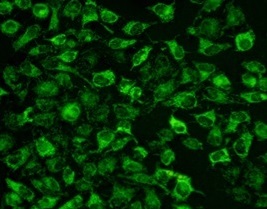
Fig. Hela cells stained with TraKine™ Mitochondrion Staining Kit (Green Fluorescence).
![]() TraKine™粒腺體染色試劑盒(Mitochondrion Staining Kits, KTC4003)是一套螢光成像工具,可標記活細胞的粒線體(mitochondria)。該試劑盒使用專有的粒線體綠色螢光探針(mitochondrial green fluorescent probe, Ex / Em = 490/523 nm),與其他探針不同,該探針似乎定位於粒線體(mitochondria),取決於粒線體膜電位(mitochondrial membrane potential)的情況要少得多。該染料是疏水性化合物(hydrophobic compound),具有額外的優勢,即它在水溶液中基本不發螢光,並且僅在粒線體的脂質(lipid environment)環境中積累後才發螢光,並且可以染色活細胞(live cells)和甲醛(formaldehyde-fixed cells)固定的細胞,但在滲透(permeabilized)後,螢光信號將丟失。可以使用螢光成像,高含量成像,微板螢光法(microplate fluorometry)或流式細胞術(flow cytometry)來測量螢光。該試劑盒適用於增殖(proliferating)和非增殖細胞(non-proliferating cells),可用於懸浮細胞(suspension)和貼壁細胞(adherent cells)。
TraKine™粒腺體染色試劑盒(Mitochondrion Staining Kits, KTC4003)是一套螢光成像工具,可標記活細胞的粒線體(mitochondria)。該試劑盒使用專有的粒線體綠色螢光探針(mitochondrial green fluorescent probe, Ex / Em = 490/523 nm),與其他探針不同,該探針似乎定位於粒線體(mitochondria),取決於粒線體膜電位(mitochondrial membrane potential)的情況要少得多。該染料是疏水性化合物(hydrophobic compound),具有額外的優勢,即它在水溶液中基本不發螢光,並且僅在粒線體的脂質(lipid environment)環境中積累後才發螢光,並且可以染色活細胞(live cells)和甲醛(formaldehyde-fixed cells)固定的細胞,但在滲透(permeabilized)後,螢光信號將丟失。可以使用螢光成像,高含量成像,微板螢光法(microplate fluorometry)或流式細胞術(flow cytometry)來測量螢光。該試劑盒適用於增殖(proliferating)和非增殖細胞(non-proliferating cells),可用於懸浮細胞(suspension)和貼壁細胞(adherent cells)。
- Kit components
- MitoGreen™ (1000×)
• Assay Buffer (10×) - Features & Benefits
- Enables the uniform staining of mitochondrion across a wide variety of mammalian cell types, and can be used for both living and fixed suspended or attached cells.
• Optimized for various fluorescence platforms such as microplate assays, flow cytometry and fluorescence microscope.
• Proprietary MitoGreen™ (Ex/Em = 490/523 nm)-much less depending on mitochondrial membrane potential. - Storage instructions
Refer to list of materials supplied for storage conditions of individual components. Stable for at least 6 months at recommended temperature from date of shipment.
![]()
| 產品貨號 | 產品品項 | 反應 | 測量儀器 |
|
|
TUNEL細胞凋亡檢測試劑盒 | 50個反應 | 螢光酶標儀、螢光顯微鏡或流式細胞儀。FITC通道(Ex / Em = 490nm / 520 nm) |
| 貨號KTA0002 | Annexin V-AbFluor™488細胞凋亡檢測試劑盒 | 50個反應 | Annexin V- AbFluor™ 488: Abs/Em = 491/517 nm; PI: Abs/Em = 535/617 nm (with DNA) |
| 貨號KTA4001 | JC-1線粒體膜電位測定試劑盒 | 20個反應 |
(聚結的JC-1,Ex / Em = 585/590 nm)發出綠色螢光(JC-1單體,Ex / Em = 510/527 nm) JC-1 Stain CCCP (10 mM) Assay Buffer (5×) |
| 貨號BMD00063 | DAPI Staining Solution | 10MG | λEx/λEm: 358/461 nm (with DNA) |
| KTD102-EN | 凋亡細胞分析(Annexin V法和TUNEL法) | 40個分析/20個分析 | |
| 貨號KTC4100 | Pro活細胞微管蛋白染色 | 50個反應/250個反應 | Ex/Em = 500/520 nm |
| 貨號KTC4001 | Abbkine 細胞膜染色 | 100個反應/500個反應 | Ex/Em = 484/501 nm |
| 貨號KTC4003 | TraKine™粒線體染色 | 100個反應 | Ex / Em = 490/523 nm增殖(proliferating)和非增殖細胞(non-proliferating cells) |