【細胞增殖&毒理&衰老】Abbkine促銷優惠活動DM&產品資料
【細胞增殖&毒理&衰老】| DNA Damege損傷/細胞Stress | 細胞凋亡&細胞染色 | 發炎反應ELISA分析 | 膽固醇代謝/代謝試驗 | 生化檢測試劑盒 | 抗體蛋白純化&其它 | Tag 標籤單多株抗體

![]()
活細胞和死細胞雙染色套組 | Live and Dead Cell Double Staining Kit貨號KTA1001
Detect using either microscopy or FACS.
![]()
活細胞和死細胞數量的定量是細胞生物學研究中不可缺少的工具。死細胞對於研究生長控制和細胞死亡非常重要。Abbkine 的活/死細胞可行性測定試劑盒 (KTA1001)提供了一種雙色螢光方法,該方法基於使用兩種不同的染料同時測定活細胞和死細胞。
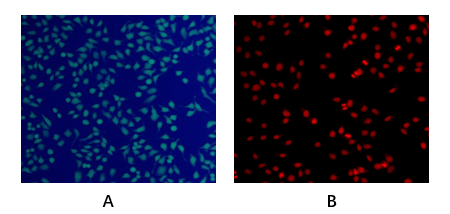
![]() Abbkine 活細胞和死細胞雙染色試劑盒 (Live and Dead Cell Double Staining Kit, KTA1001) 提供了一個方便的測定評估死活細胞的快速方法,基於同時測定活細胞和死細胞與兩種Probe,測量細胞健康、血漿膜完整性、和細胞內酯酶活性。該試劑盒利用LiveDye,一種細胞滲透的綠色螢光染料(Ex/Em = 488/530 nm),染色活細胞和核染料,一種細胞不可滲透的紅色螢光染料(Ex/Em = 535/617)以染色死細胞。
Abbkine 活細胞和死細胞雙染色試劑盒 (Live and Dead Cell Double Staining Kit, KTA1001) 提供了一個方便的測定評估死活細胞的快速方法,基於同時測定活細胞和死細胞與兩種Probe,測量細胞健康、血漿膜完整性、和細胞內酯酶活性。該試劑盒利用LiveDye,一種細胞滲透的綠色螢光染料(Ex/Em = 488/530 nm),染色活細胞和核染料,一種細胞不可滲透的紅色螢光染料(Ex/Em = 535/617)以染色死細胞。
![]()
1. Ocular surface repair using decellularized porcine conjunctiva
L Zhao, Y Jia, C Zhao. Acta biomaterialia, 2019 – Elsevier
2. Grafting antibacterial polymer brushes from titanium surface via polydopamine chemistry and activators regenerated by electron transfer ATRP
Shuangshuang Wang, Jixuan Song, Yuchao Li, et al. Reactive and Functional Polymers. July 2019, 140: 48-55.
細胞衰老檢測試劑盒(β-半乳糖苷酶) | 貨號KTA3030
Cell Senescence Kit (β-Galactosidase Assay)
![]() 細胞衰老(Cellular senescence)是正常細胞停止分裂(cell divide)的現象。衰老的細胞(senescent cells)不再能夠複製(replicate),但它們仍保持代謝活性(metabolically active),但因阻滯於G1期,失去了對有絲分裂原的反應能力和合成DNA的能力,不能進入S期。並且與衰老相關的β-半乳糖苷酶活性染色(senescence-associated beta-galactosidase activity)呈陽性,這被認為是細胞衰老的生物標記(biomarker)。細胞的衰老(Cellular senescence)可以通過激活癌基因(oncogenes)和細胞-細胞融合(cell-cell fusion)來實現,DNA對活性氧(reactive oxygen species , ROS)升高的反應會導致DNA損傷(DNA damage),而不是僅僅依賴於細胞分裂的數目。在正常衰老過程(normal aging)中,組織中的衰老細胞數量顯著增加。
細胞衰老(Cellular senescence)是正常細胞停止分裂(cell divide)的現象。衰老的細胞(senescent cells)不再能夠複製(replicate),但它們仍保持代謝活性(metabolically active),但因阻滯於G1期,失去了對有絲分裂原的反應能力和合成DNA的能力,不能進入S期。並且與衰老相關的β-半乳糖苷酶活性染色(senescence-associated beta-galactosidase activity)呈陽性,這被認為是細胞衰老的生物標記(biomarker)。細胞的衰老(Cellular senescence)可以通過激活癌基因(oncogenes)和細胞-細胞融合(cell-cell fusion)來實現,DNA對活性氧(reactive oxygen species , ROS)升高的反應會導致DNA損傷(DNA damage),而不是僅僅依賴於細胞分裂的數目。在正常衰老過程(normal aging)中,組織中的衰老細胞數量顯著增加。

細胞是生物結構(biological structure)和功能(function)的基本單位,也是生物衰老(biological aging)的基本單位。細胞衰老(Cellular senescence)在形態上表現為細胞結構(cell structure)的退化,例如核膜凹陷(nuclear membrane depression),最終導致核膜塌陷(nuclear membrane collapse),染色質結構(chromatin structure)改變,超二倍體(hyperdiploid)和異常多倍體細胞(polyploid cells)數量增加。細胞膜的脆性(cell membrane fragility)增加。通透性(permeability)降低,膜受體(membrane receptors)的類型和數量以及對配體(ligands)的敏感性發生變化;脂褐素(lipofuscin)在細胞中積累,許多細胞器和細胞內結構發生變性。細胞衰老(Cell senescence)在生理上表現(physiologically manifested)為功能下降和新陳代謝低下,例如細胞週期停滯(cell cycle arrest),細胞複製能力 (cell replication ability) 喪失,對促有絲分裂刺激(mitogenic stimulation)的反應性減弱以及對促凋亡因子(pro-apoptotic factors)的反應性改變。細胞內酶活性中心被氧化,酶活性(Enzyme activity)降低,蛋白質合成(protein synthesis)降低等。衰老細胞(senescent cells)不再複制,但它們仍保持代謝活性(metabolically active),並且衰老相關的β-半乳糖苷酶活性(beta-galactosidase activity)呈陽性,這被認為是細胞衰老生物標記(biomarker of cellular senescence)。
![]()
產品描述: Abbkine細胞衰老檢測試劑盒(Senescence β-Galactosidase Staining Kit, KTA3030)提供了基於衰老過程中pH值為6的衰老相關β-半乳糖苷酶(SA-β-Gal)活性上調的試劑,用於檢測衰老細胞(senescent cells)或組織(tissues)。細胞或組織的老化可以在普通的光學顯微鏡下觀察。 SA-β-Gal僅存在於衰老細胞(senescent cells)中,在衰老前,靜止,腫瘤或永生細胞中無法檢測到。
Senescence β-Galactosidase Staining Kit provide reagents for detecting the senescent cells or tissues based on the up-regulation of senescence-associated β-galactosidase (SA-β-Gal) activity at pH 6 during aging. The aging of cells or tissues can be observed under a common optical microscope. SA-β-Gal is present only in senescent cells and is undetectable in presenescent, quiescent, tumor or immortal cells
- Kit components
- 10×Fixation Buffer • 10×PBS • Reagent A • Reagent B • Reagent C • X-Gal solution
- Features & Benefits
- Simple and optimized procedure. • Fast and convenient. • Suitable for aging detection of cultured cells and tissue sections. • Cell senescence β-galactosidase staining kit only stains senescent cells, and does not stain pre-senescence cells (senescent cells), quiescent cells (static cells), immortal cells (immortal cells) or tumor cells.
- Storage instructions
Refer to list of materials supplied for storage conditions of individual components. Stable for at least 12 months at recommended temperature from date of shipment.
- Shipping
Gel pack with blue ice.
- Precautions
The product listed herein is for research use only and is not intended for use in human or clinical diagnosis. Suggested applications of our products are not recommendations to use our products in violation of any patent or as a license. We cannot be responsible for patent infringements or other violations that may occur with the use of this product.
CCK-8細胞增殖和細胞毒性試劑盒 | 貨號 KTA1020 吸收光450 nm CCK8操作手冊
![]()
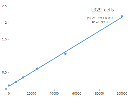
Fig. Abbkine Economical Cell Counting Kit-8 (CCK-8) is designed to detect cell proliferation and cell toxicity.
CCK-8; WST-8; CCK8; WST8
![]() Abbkine Cell Counting Kit-8(CCK-8)設計用於檢測細胞增殖和細胞毒性。細胞計數試劑盒-8(CCK-8)使用高度水溶性的四唑鹽WST-8:[2-(2-甲氧基-4-硝基苯基)-3-(4-硝基苯基)-5-(2,4-二磺基苯基 )-2H-四唑鎓單鈉鹽]通過還原轉化為水溶性橙色甲dye染料。 CCK-8具有低毒性,並且允許用於測定細胞增殖和細胞毒性測定中活細胞數量的靈敏比色測定。 細胞中脫氫酶產生的甲dye染料的量與活細胞的數量成正比。Cell Counting Kit-8(CCK-8)用於檢測細胞增殖(cell proliferation)及細胞毒性(cell toxicity),其原理基於WST-8經細胞中的脫氫酶降低,得到橙色產物(formazan)。細胞中formazan的含量與活細胞數成正比。細胞計數試劑盒8(Cell Counting Kit-8, CCK-8)通過使用高度水溶性的四唑鹽-WST-8進行非常方便的測定。[2-(2-甲氧基-4-硝基苯基)-3-(4-硝基苯基)-5- (2,4-二磺基苯基)-2H-四唑鎓鹽]在電子介體的存在下還原生成水溶性formazan dye染料。 CCK-8是非放射性的,可進行敏感的比色測定,以測定細胞增殖(cell proliferation)和細胞毒性測定中的活細胞數量。 WST-8被細胞中的脫氫酶(dehydrogenases)還原,得到橙色產物(formazan),可溶於組織培養基。細胞中脫氫酶(dehydrogenases) 產生的formazan dye染料的量與活細胞的數量成正比。WST-8生產的產品(formazan)是水溶性的,不需要有機溶劑或同位素。而且formazan是穩定和安全的。使用CCK-8的檢測靈敏度高於使用其他四唑鹽(tetrazolium salts, 如MTT,XTT,MTS或WST-1)的檢測靈敏度。
Abbkine Cell Counting Kit-8(CCK-8)設計用於檢測細胞增殖和細胞毒性。細胞計數試劑盒-8(CCK-8)使用高度水溶性的四唑鹽WST-8:[2-(2-甲氧基-4-硝基苯基)-3-(4-硝基苯基)-5-(2,4-二磺基苯基 )-2H-四唑鎓單鈉鹽]通過還原轉化為水溶性橙色甲dye染料。 CCK-8具有低毒性,並且允許用於測定細胞增殖和細胞毒性測定中活細胞數量的靈敏比色測定。 細胞中脫氫酶產生的甲dye染料的量與活細胞的數量成正比。Cell Counting Kit-8(CCK-8)用於檢測細胞增殖(cell proliferation)及細胞毒性(cell toxicity),其原理基於WST-8經細胞中的脫氫酶降低,得到橙色產物(formazan)。細胞中formazan的含量與活細胞數成正比。細胞計數試劑盒8(Cell Counting Kit-8, CCK-8)通過使用高度水溶性的四唑鹽-WST-8進行非常方便的測定。[2-(2-甲氧基-4-硝基苯基)-3-(4-硝基苯基)-5- (2,4-二磺基苯基)-2H-四唑鎓鹽]在電子介體的存在下還原生成水溶性formazan dye染料。 CCK-8是非放射性的,可進行敏感的比色測定,以測定細胞增殖(cell proliferation)和細胞毒性測定中的活細胞數量。 WST-8被細胞中的脫氫酶(dehydrogenases)還原,得到橙色產物(formazan),可溶於組織培養基。細胞中脫氫酶(dehydrogenases) 產生的formazan dye染料的量與活細胞的數量成正比。WST-8生產的產品(formazan)是水溶性的,不需要有機溶劑或同位素。而且formazan是穩定和安全的。使用CCK-8的檢測靈敏度高於使用其他四唑鹽(tetrazolium salts, 如MTT,XTT,MTS或WST-1)的檢測靈敏度。
- Formulation
Liquid solution
- Kit components
- CCK-8 solution
- Features & Benefits
- One-bottle, ready-to-use solution.
• The product (formazan) produced by WST-8 is water soluble, No organic solvents or isotopes required. And the formazan is stable and safe.
• The detection sensitivity using CCK-8 is higher than assays using other tetrazolium salts such as MTT, XTT, MTS or WST-1.
• The same cells can be used for other cell assays because of the low toxicity of CCK-8. - Storage instructions
CCK-8 is stable over one year at 0-5°C with protection from light. Store it at -20°C for longer storage. Repeated thawing and freezing causes an increase in the background, which interferes with the assay. Please store the kit at 0-5°C for frequent use.
細胞增殖EdU Image試劑盒 | Cell Proliferation EdU Image Kit貨號KTA2030
【DNA合成/細胞增殖檢測】 EdU Image試劑盒-BrdU分析新型替代方法
細胞增殖(cell proliferation)的檢測,對於評估細胞健康(cell health),確定遺傳毒性(genotoxicity)或評估抗癌藥物(anticancer drugs)至關重要。到目前為止,直接測量DNA合成(DNA synthesis)是最準確的方法,通常是通過在複製過程中將[3H]胸苷(thymidine)或5-溴-2′-脫氧尿苷(5-bromo-2’-deoxyuridine)等核苷類似物(nucleoside analog like)摻入細胞中,然後通過放射自顯影或分別使用抗BrdU抗體(anti-BrdU-antibody)。EdU(5-乙炔基-2′-脫氧尿苷)是BrdU(5-bromo-2′-脫氧尿苷)的新型替代品。它被取代胸苷摻入到新合成的增殖細胞DNA中。將5-EdU摻入到增殖細胞中,然後使用點擊連接用螢光疊氮化物進行檢測。與BrdU的使用相反,新方法不需要樣品固定或DNA變性。可用於研究中樞神經系統中的細胞增殖。
細胞增殖EdU Image試劑盒 | Cell Proliferation EdU Image Kit貨號KTA2030

產品描述:
細胞增殖EdU Image試劑盒(Cell Proliferation EdU Image Kit, 貨號KTA2030)(綠色螢光)是BrdU分析的一種新型替代方法。 EdU(5-乙炔基-2′-脫氧尿苷)是胸苷的核苷類似物,在活性DNA合成過程中被摻入DNA中。與BrdU分析相比,EdU-Click分析不是基於抗體的,因此不需要DNA變性(denaturation)(通常使用HCl或加熱或用DNase消化)來檢測摻入的核苷。檢測基於click reaction,疊氮化物和炔烴之間的銅催(copper-catalyzed)化共價反應在30分鐘內完成。
| Product name | EdU Cell Proliferation Image Kit (Green Fluorescence) |
| Kit components | • EdU (10mM) • AbFluor 488 azide • 10×Reaction buffer • Copper • Reducing Agent |
| Features & Benefits | • No antibody needed. • No denaturation steps, preservation of cell morphology and DNA integrity. • Simple, reliable and time-saving than traditional methods. • Proprietary AbFluor 488 azide (Ex/Em = 501/525 nm)-good photostability and minimizing fluorescence quenching. • Optimized for fluorescent microscopy. |
| Storage instructions | Stable for at least 12 months at recommended temperature from date of shipment. Gel pack with blue ice. |
| Shipping | Gel pack with blue ice. |
| Background | The detection of cell proliferation is of utmost importance for assessing cell health, determining genotoxicity or evaluating anticancer drugs. Until now, measuring DNA synthesis directly is most accurate method of doing it, normally performed by incorporation of the nucleoside analog like [3H] thymidine or 5-bromo-2’-deoxyuridine to cells during replication, and then detected or visualized by autoradiography or with an anti-BrdU-antibody respectively. |
引用文獻
In vitro mitochondrial-targeted antioxidant peptide induces apoptosis in cancer cells. W Zhan, X Liao, L Li. Onco Targets Therv.12
Perilaldehyde activates AMP‐activated protein kinase to suppress the growth of gastric cancer via induction of autophagy
Y Zhang, S Liu, Q Feng. JOURNAL OF CELLULAR BIOCHEMISTRY
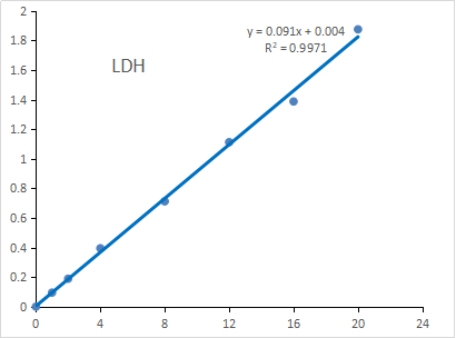
乳酸脫氫酶(LDH)測定試劑盒 | 貨號KTB1110
The kit can detect 1- 20 U/mL of LDH directly in samples
![]() 乳酸脫氫酶(Lactate dehydrogenase , LDH)是一種存在於多種生物體中的氧化還原酶(oxidoreductase)。LDH催化丙酮酸鹽(pyruvate)和乳酸鹽(lactate)的相互轉化,伴隨著NADH和NAD+的相互轉化。LDH在缺氧條件下將糖酵解(glycolysis)的最終產物丙酮酸轉化為乳酸(lactate)。乳酸脫氫酶定量具有臨床意義,因為某些乳酸脫氫酶同工酶的血清水準反映了特定組織的病理狀況。當疾病、損傷或有毒物質損壞組織時,細胞乳酸脫氫酶釋放到血流中。由於乳酸脫氫酶是一種相當穩定的酶,乳酸脫氫酶已被廣泛用於評估組織和細胞的損傷和毒性。CheKine™乳酸脫氫酶測定試劑盒(Lactate Dehydrogenase Assay Kit, KTB1110) 提供了一種簡單而簡便的比色測定法,用於測量血清,血漿,細胞培養上清液,組織/細胞裂解液培養基,發酵液和其他生物液體中的乳酸脫氫酶(Lactate Dehydrogenase)。 在此比色測定中,LDH將NAD還原為NADH,然後與探針相互作用以產生顏色(λmax= 450 nm)。該測定快速且方便。
乳酸脫氫酶(Lactate dehydrogenase , LDH)是一種存在於多種生物體中的氧化還原酶(oxidoreductase)。LDH催化丙酮酸鹽(pyruvate)和乳酸鹽(lactate)的相互轉化,伴隨著NADH和NAD+的相互轉化。LDH在缺氧條件下將糖酵解(glycolysis)的最終產物丙酮酸轉化為乳酸(lactate)。乳酸脫氫酶定量具有臨床意義,因為某些乳酸脫氫酶同工酶的血清水準反映了特定組織的病理狀況。當疾病、損傷或有毒物質損壞組織時,細胞乳酸脫氫酶釋放到血流中。由於乳酸脫氫酶是一種相當穩定的酶,乳酸脫氫酶已被廣泛用於評估組織和細胞的損傷和毒性。CheKine™乳酸脫氫酶測定試劑盒(Lactate Dehydrogenase Assay Kit, KTB1110) 提供了一種簡單而簡便的比色測定法,用於測量血清,血漿,細胞培養上清液,組織/細胞裂解液培養基,發酵液和其他生物液體中的乳酸脫氫酶(Lactate Dehydrogenase)。 在此比色測定中,LDH將NAD還原為NADH,然後與探針相互作用以產生顏色(λmax= 450 nm)。該測定快速且方便。
![]()
| 產品名稱 | 貨號 |
| CheKine™ Lactate Dehydrogenase Assay Kit 乳酸脫氫酶分析試劑盒 |
KTB1110 |
原理:為定量樣本中乳酸脫氫酶(LDH)提供了一個簡單便捷的比色法方案。乳酸在乳酸脫氫酶(LDH)的作用下生成丙酮酸,同時使NAD+還原生成NADH,NADH還原WST-8生成橙黃色物質,在450nm處的OD值與LDH成正比。
適用樣本:血清、血漿、細胞培養上清、組織/細胞裂解物、發酵液及其它生物液體。
| Kit components | • Assay Buffer • Lactate • NAD • WST-8 • Enhancer • Lactate Dehydrogenase Standard (100 U/mL) |
| Features & Benefits | • Optimized procedure for measuring Lactate Dehydrogenase (LDH) in various samples containing serum, plasma, cell culture supernatants, tissue/cell lysates culture medium, fermentation and other biological fluids. • Simple and easy colorimetric assay. • Quick and convenient. |
| Calibration range | 1- 20 U/mL |
| Usage notes | • Briefly centrifuge small vials at low speed prior to opening. • Performing several dilutions of your sample to ensure the readings are within the standard value range. • Use fresh samples. If not assayed immediately, samples can be stored at -80°C for one month. |
| Storage instructions | Storage at –20°C and Keep from light. Stable for at least 6 months at recommended temperature from date of shipment. |
| Shipping | Gel pack with blue ice. |
細胞週期染色檢測 | Cell Cycle Staining Kit貨號 KTA2020
細胞週期染色 (Cell Cycle) 研究的意義
細胞DNA含量的測量,是確定細胞週期階段(cell cycle)(G1,S和/或G2 / M)的最常見方法。是基於隨著細胞在細胞週期中,G1期的細胞具有一組成對的染色體,並且在DNA含量方面是一致的。 另一方面,DNA的數量在S期開始增加一倍,因此DNA的數量是G1數量的一到兩倍。 與G1中的細胞和兩組成對的染色體相比,G2 / M期的細胞具有的DNA量是其兩倍。Abbkine細胞週期染色試劑盒(Cell Cycle Staining Kit, KTA2020) 利用一種核染料(nuclear dye),該核染料與細胞中的核酸結合會產生螢光信號(fluorescence signal),該信號與細胞DNA含量(DNA content) 成正比,可方便,準確地確定細胞各相中細胞週期(cell cycle)的百分比。
![]()
| Product name | Cell Cycle Staining Kit |
| Applications notes | Abbkine Cell Cycle Staining Kit utilizes a nuclear dye, the binding of which to nucleic acids in the cell results in fluorescence signal, which is proportional to cellular DNA content, providing a convenient and accurate determination of the percentage of cells in each phase of the cell cycle. |
| Kit components | • Nuclear Dye (50×) • Assay Buffer (10×) • RNase A (100×) |
| Features & Benefits | • Flow cytometric analysis of cell cycle progression. • Ready-to-use reagents for staining cells. • The appropriate observe channel is Ex/Em=535/615 nm. |
| Usage notes | The optimal concentration of the Nuclear Dye and incubation time varies depending on the specific application. The staining conditions may need modified according to the particular cell type. |
| Storage instructions | Stable for at least 12 months at recommended temperature from date of shipment. Gel pack with blue ice. |
| Shipping | Gel pack with blue ice. |
| Background | The most common approach to determining the cell cycle stage (G1, S, and/or G2/M) is based on measurement of cellular DNA content. As cells progress through the cell cycle, cells in the G1 phase have one set of paired chromosomes and are uniform with respect to DNA content. On the other hand, the amount of DNA begins to double during S phase, so that the amount of DNA is between one and two times the amount in G1. Cells in G2/M phase have double the amount of DNA compared to cells in G1 and two sets of paired chromosomes. |
LDH 細胞毒性檢測試劑盒 | LDH Cytotoxicity Assay Kit貨號KTA1030
LDH Cytotoxicity Assay Kit
細胞死亡(Cell death)或細胞毒性(cytotoxicity)通常通過質膜損傷(plasma membrane damage)的量化來評估。 LDH 是一種穩定的酶,存在於所有細胞類型中,並在質膜(plasma membrane)受損後迅速釋放到細胞培養基中。 因此,LDH 是細胞毒性(cytotoxicity)研究中使用最廣泛的標誌物。
![]() 細胞毒性檢測試劑盒(Abbkine LDH Cytotoxicity Assay Kit, KTB1030)為細胞毒性(cytotoxicity)研究提供了一種簡單易行的比色檢測方法(colorimetric assay)。 該測定基於在 NADH 偶聯酶促反應(NADH-coupled enzymatic reaction)中將四唑鹽 MTT (tetrazolium salt MTT) 還原為 MTT 的還原形式,其在 565 nm 處具有最大吸收。 生成顏色的強度與裂解的細胞數量直接相關。 在典型的細胞毒性試驗(typical cytotoxicity assay)中,靶細胞(target cells)與細胞毒性化學試劑(cytotoxic chemical agent)或細胞毒性細胞(NK 細胞、細胞毒性 T 細胞)一起培養,以誘導靶細胞 (target cells)死亡和 LDH 釋放。 將含有 LDH 的上清液轉移到新的 96 孔測定板的孔中,並與 LDH 反應溶液混合。 在室溫下孵育 30 分鐘後,使用讀板器讀取 565nm (A565) 處的吸光度。
細胞毒性檢測試劑盒(Abbkine LDH Cytotoxicity Assay Kit, KTB1030)為細胞毒性(cytotoxicity)研究提供了一種簡單易行的比色檢測方法(colorimetric assay)。 該測定基於在 NADH 偶聯酶促反應(NADH-coupled enzymatic reaction)中將四唑鹽 MTT (tetrazolium salt MTT) 還原為 MTT 的還原形式,其在 565 nm 處具有最大吸收。 生成顏色的強度與裂解的細胞數量直接相關。 在典型的細胞毒性試驗(typical cytotoxicity assay)中,靶細胞(target cells)與細胞毒性化學試劑(cytotoxic chemical agent)或細胞毒性細胞(NK 細胞、細胞毒性 T 細胞)一起培養,以誘導靶細胞 (target cells)死亡和 LDH 釋放。 將含有 LDH 的上清液轉移到新的 96 孔測定板的孔中,並與 LDH 反應溶液混合。 在室溫下孵育 30 分鐘後,使用讀板器讀取 565nm (A565) 處的吸光度。
![]()
Abbkine LDH Cytotoxicity Assay Kit provides a simple and easy colorimetric assay for the study of cytotoxicity. This assay is based on the reduction of the tetrazolium salt MTT in a NADH-coupled enzymatic reaction to a reduced form of MTT which exhibits an absorption maximum at 565 nm. The intensity of the generated color correlates directly with the cell number lysed. In a typical cytotoxicity assay, target cells are cultured with a cytotoxic chemical agent or a cytotoxic cell (NK cell, cytotoxic T cells) to induce target cell death and LDH release. The LDH-containing supernatants are transferred to wells of a new 96-well assay plate and mixed with the LDH Reaction solution. After an incubation of 30 minutes at room temperature, the absorbance at 565nm (A565) is read using a plate reader. Abbkine LDH
| Kit components | • Assay Buffer • Lactic Acid Solution • MTT Solution • PES Solution • LDH Positive Control • NAD+ Solution • Triton X-100 (10%) |
| Features & Benefits | • Measures cell death in response to chemical compounds or environmental factors. • LDH release from the cytosol into the medium catalyzes a color-forming reaction. • Detect lysis of 10,00-100,0000 cells per well. |
| Usage notes | Performing an initial titration experiment to determine the optimal number of cells per well of the target cell you plan to use. |
| Storage instructions | Store at -20°C and Keep in dark. The Kit has a storage time of 6 months from receipt. |
| Shipping | Gel pack with blue ice. |
| Precautions | The product listed herein is for research use only and is not intended for use in human or clinical diagnosis. Suggested applications of our products are not recommendations to use our products in violation of any patent or as a license. We cannot be responsible for patent infringements or other violations that may occur with the use of this product. |
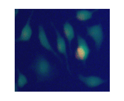
Calcein AM鈣黃綠素活細胞螢光染劑 | CAS#148504-34-1貨號BMD00064-50UG 操作手冊

![]() 鈣黃綠素AM(Calcein AM) 是一種細胞可滲透(cell-permeable)的染料,常用於活細胞螢光標記,Calcein(鈣黃綠素)的基礎上加強了疏水性,因此能夠輕易穿透活細胞膜。當其進入到活細胞(live cells)後,會被細胞內酯酶(intracellular esterases)裂解,留下膜不滲透(membrane-impermeant)的鈣黃綠素(Calcein),Calcein這是一種螢光指示劑,發出強綠色螢光。其最大吸收和發射波長分別為494和517 nm。它可用作細胞活力(cell viability),細胞間通訊(cell-cell communication),細胞毒性(cytotoxicity)或細胞內鈣(calcium),氟(fluoride),鐵(iron)或汞(mercury)變化的指標。由於鈣黃綠素是多藥抗性相關蛋白1(multidrug resistance-associated protein 1, MRP1)和多藥抗性蛋白3(multidrug resistance protein 3, MDR3,P-糖蛋白3)的底物,因此鈣黃綠素AM(Calcein AM)用於研究這些蛋白及其調節。
鈣黃綠素AM(Calcein AM) 是一種細胞可滲透(cell-permeable)的染料,常用於活細胞螢光標記,Calcein(鈣黃綠素)的基礎上加強了疏水性,因此能夠輕易穿透活細胞膜。當其進入到活細胞(live cells)後,會被細胞內酯酶(intracellular esterases)裂解,留下膜不滲透(membrane-impermeant)的鈣黃綠素(Calcein),Calcein這是一種螢光指示劑,發出強綠色螢光。其最大吸收和發射波長分別為494和517 nm。它可用作細胞活力(cell viability),細胞間通訊(cell-cell communication),細胞毒性(cytotoxicity)或細胞內鈣(calcium),氟(fluoride),鐵(iron)或汞(mercury)變化的指標。由於鈣黃綠素是多藥抗性相關蛋白1(multidrug resistance-associated protein 1, MRP1)和多藥抗性蛋白3(multidrug resistance protein 3, MDR3,P-糖蛋白3)的底物,因此鈣黃綠素AM(Calcein AM)用於研究這些蛋白及其調節。
![]() Calcein AM 活細胞螢光染劑 | 用於細胞活性分析之建議配製
Calcein AM 活細胞螢光染劑 | 用於細胞活性分析之建議配製
Calcein AM分子量為995 g/mole , 稱取50ug的calcein AM可以溶在50ul的DMSO ;最後濃度為1Mm取1ul的Calcein AM 溶在1X 的1000ul dissociation solution =1uM (1000ul)以96well 為例,加入100ul的1uM Calcein AM,大約可以使用500次。Calcein-AM的ester部位遇到濕氣會分解,使用後請在-20℃下密閉保存,溶於DMSO後建議2個月內使用完。
![]()
| Product name | Calcein AM |
| CAS Number | 148504-34-1 |
| Molecular Formula | C46H46N2O23 |
| Alternative | Calcein AM,BMD00064 |
| Formulation | Gray solid |
| Features & Benefits | • λEX/λEm: 494/517 nm (pH 8) (calcein) |
| Molecular weight | 995 |
| Usage notes | • Soluble in DMSO |
| Storage instructions | Store at -20°C. The product can be stored for up to 12 months. Protect from light. |
| Shipping | Gel pack with blue ice. |
| 產品貨號 | 產品品項 | 反應 | 測量儀器 |
| 貨號KTA1001 | 活&死細胞雙染色試劑盒 | 100個反應 |
活細胞綠色螢光染劑Ex/Em = 488/530 nm; 死細胞紅色螢光染料(Ex/Em = 535/617) |
| 貨號KTA3030 | 細胞衰老檢測(β-半乳糖苷酶) | 100個反應 | 光學顯微鏡 |
| 貨號KTA1020 | CCK-8細胞增殖和細胞毒性試劑盒 | 1000個反應 | 吸收光450nm |
| 貨號KTA2030 | EdU Image細胞增殖試劑盒 (BrdU替代法) | 100個反應 | AbFluor 488 azide (Ex/Em = 501/525 nm) |
| 貨號KTB1110 | (LDH)乳酸脫氫酶測定試劑盒 | 96個反應/ |
λmax= 450 nm 1- 20 U/mL of LDH |
| 貨號 KTA2020 | 細胞週期染色檢測 | 50個反應 | Ex/Em=535/615 nm. |
| 貨號KTA1030 | LDH細胞毒性測定試劑盒 | 96個反應 |
OD 565 nm 10,00-100,0000 cells per well |
| 貨號BMD00064-50UG | Calcein AM鈣黃綠素活細胞螢光染劑 | 50ug |
λEX/λEm: 494/517 nm (pH 8) (calcein)
|