Ki-67單株抗體 | 貨號ABM40064

| 代理廠牌: | |
| 原廠連結: | |
| 相關下載: |
細胞增殖(Cell proliferation)可用於評估正常的細胞健康狀況(normal cell health),測量對毒性損傷(toxic insult)的反應,或用作幾種癌症的預後(prognostic)和診斷工具(diagnostic tool)。可用的標記通常著重於DNA水平(DNA levels)或合成(synthesis),細胞代謝(cellular metabolism)或增殖特異性蛋白(proliferation-specific proteins)。
產品描述: Ki-67是一種由人MKI67基因編碼的蛋白質。該蛋白與細胞的增殖密切相關。MKI67編碼核蛋白(nuclear protein)(增殖Ki-67的標記)與細胞增殖(cellular proliferation)相關。Alternatively spliced transcript variants have been described. A related pseudogene exists on chromosome X.
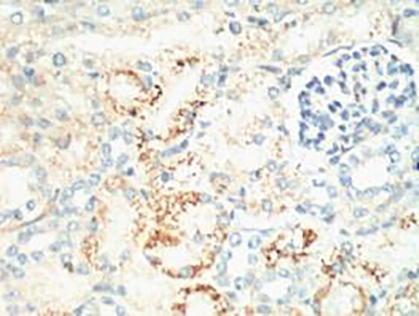
Fig.1. IHC-P staining of mouse kidney tissue, diluted at 1:200.
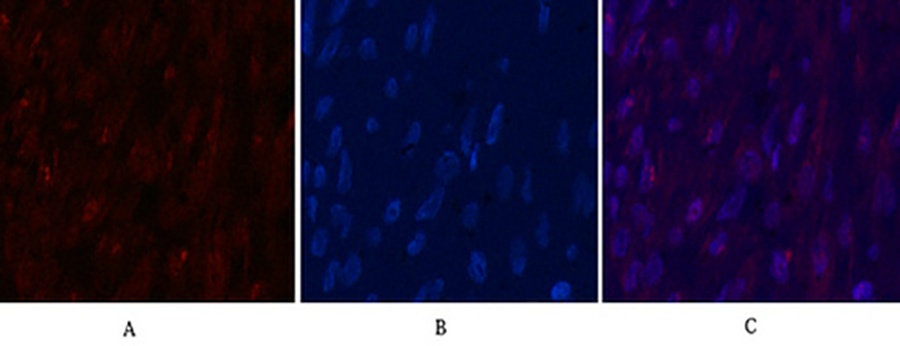
Fig.2. Immunofluorescence analysis of human breast cancer tissue. 1, Ki 67 Monoclonal Antibody (red) was diluted at 1:200 (4°C, overnight). 2, Cy3 Labeled secondary antibody was diluted at 1:300 (room temperature, 50min). 3, Picture B: DAPI (blue) 10min. Picture A: Target. Picture B: DAPI. Picture C: merge of A+B.
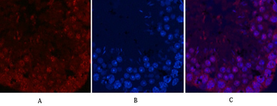
Fig.3. Immunofluorescence analysis of mouse testis tissue. 1, Ki 67 Monoclonal Antibody (red) was diluted at 1:200 (4°C, overnight). 2, Cy3 Labeled secondary antibody was diluted at 1:300 (room temperature, 50min). 3, Picture B: DAPI (blue) 10min. Picture A: Target. Picture B: DAPI. Picture C: merge of A+B.
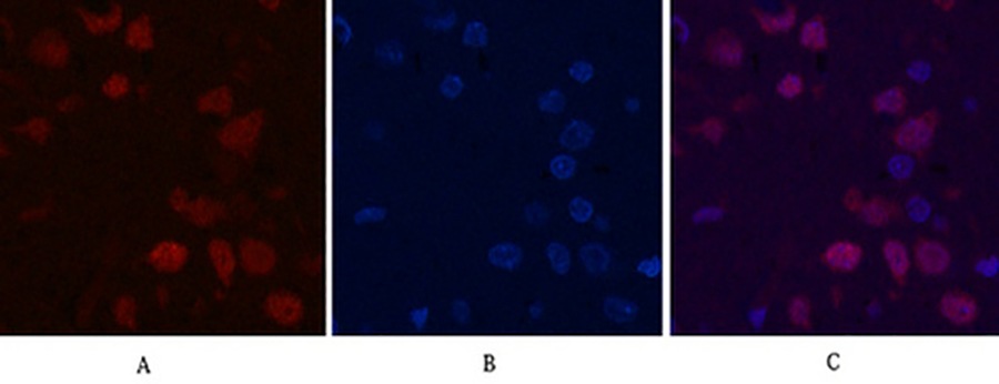
Fig.4. Immunofluorescence analysis of rat brain tissue. 1, Ki 67 Monoclonal Antibody (red) was diluted at 1:200 (4°C, overnight). 2, Cy3 Labeled secondary antibody was diluted at 1:300 (room temperature, 50min). 3, Picture B: DAPI (blue) 10min. Picture A: Target. Picture B: DAPI. Picture C: merge of A+B.
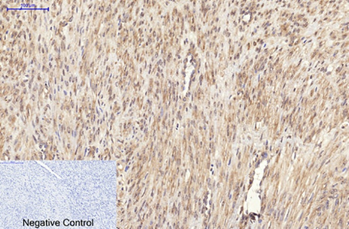
Fig.5. Immunohistochemical analysis of paraffin-embedded human uterus cancer tissue. 1, Ki 67 Monoclonal Antibody was diluted at 1:200 (4°C, overnight). 2, Sodium citrate pH 6.0 was used for antibody retrieval (>98°C, 20min). 3, secondary antibody was diluted at 1:200 (room temperature, 30min). Negative control was used by secondary antibody only.
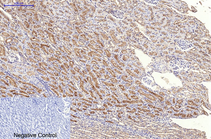
Fig.6. Immunohistochemical analysis of paraffin-embedded rat kidney tissue. 1, Ki 67 Monoclonal Antibody was diluted at 1:200 (4°C, overnight). 2, Sodium citrate pH 6.0 was used for antibody retrieval (>98°C, 20min). 3, secondary antibody was diluted at 1:200 (room temperature, 30min). Negative control was used by secondary antibody only.
- Product name
Ki 67 Monoclonal Antibody
- Immunogen
Synthetic Peptide
- Host
Mouse
- Reactivity
Human, Mouse, Rat
- Applications
IF, IHC-P
- Application notes
Optimal working dilutions should be determined experimentally by the investigator. Suggested starting dilutions are as follows: IHC-P (1:200).
- Clonality
Monoclonal
- Isotype
Mouse IgG1
- Purification
The antibody was affinity-purified from mouse ascites by affinity-chromatography using specific immunogen
- Formulation
Liquid solution
- Concentration
1 mg/ml
- Storage buffer
PBS, pH 7.4, containing 0.02% Sodium Azide as preservative and 50% Glycerol.
- Storage instructions
Stable for one year at -20°C from date of shipment. For maximum recovery of product, centrifuge the original vial after thawing and prior to removing the cap. Aliquot to avoid repeated freezing and thawing.
活細胞和死細胞雙染色套組 | CCK-8細胞增殖和細胞毒性試劑盒 | LDH細胞毒性測定試劑盒 | 細胞衰老檢測試劑盒(β-半乳糖苷酶 | 細胞增殖EdU Image試劑盒
| 貨號 | 產品名稱 | 胞器染色位置 | 螢光波段 | |
| 貨號KTC4003 | TraKine™粒線體染色 | 粒線體(mitochondria) | Ex / Em = 490/523 nm | 增殖(proliferating)和非增殖細胞(non-proliferating cells) |
| 貨號KTC4100 | Pro活細胞微管蛋白染色 | 微管蛋白(Tubulin) | Ex/Em = 500/520 nm | 活細胞或固定之細胞 |
| 貨號KTC4001 | Abbkine 細胞膜染色 | 細胞膜 (plasma membrane) | Ex/Em = 484/501 nm | 活細胞、固定懸浮、貼附細胞 |
| 貨號BMD00063 | DAPI Staining Solution | 細胞核 dsDNA | Ex/Em: 358/461 nm (with DNA) | 染色活細胞和固定細胞 |
| 貨號KTC4210 | 活細胞Lysosome染色 | Lysosome | deep red fluorescent dye , Ex/Em:650/665nm | 染色活細胞和固定細胞 |