EdU 細胞增殖試劑盒(橘) | Cell Proliferation EdU Image Kit 貨號KTA2031

| 代理廠牌: | |
| 原廠連結: | |
| 相關下載: |
【DNA合成/細胞增殖檢測】 EdU Image試劑盒-BrdU分析新型替代方法
細胞增殖(cell proliferation)的檢測,對於評估細胞健康(cell health),確定遺傳毒性(genotoxicity)或評估抗癌藥物(anticancer drugs)至關重要。到目前為止,直接測量DNA合成(DNA synthesis)是最準確的方法,通常是通過在複製過程中將[3H]胸苷(thymidine)或5-溴-2′-脫氧尿苷(5-bromo-2’-deoxyuridine)等核苷類似物(nucleoside analog like)摻入細胞中,然後通過放射自顯影或分別使用抗BrdU抗體(anti-BrdU-antibody)。EdU(5-乙炔基-2′-脫氧尿苷)是BrdU(5-bromo-2′-脫氧尿苷)的新型替代品。它被取代胸苷摻入到新合成的增殖細胞DNA中。將5-EdU摻入到增殖細胞中,然後使用點擊連接用螢光疊氮化物進行檢測。與BrdU的使用相反,新方法不需要樣品固定或DNA變性。可用於研究中樞神經系統中的細胞增殖。
EdU 摻入和檢測不需要 DNA 變性,從而減少步驟並減少組織降解。
細胞增殖EdU Image試劑盒 | Cell Proliferation EdU Image Kit (Orange Fluorescence) 貨號KTA2031
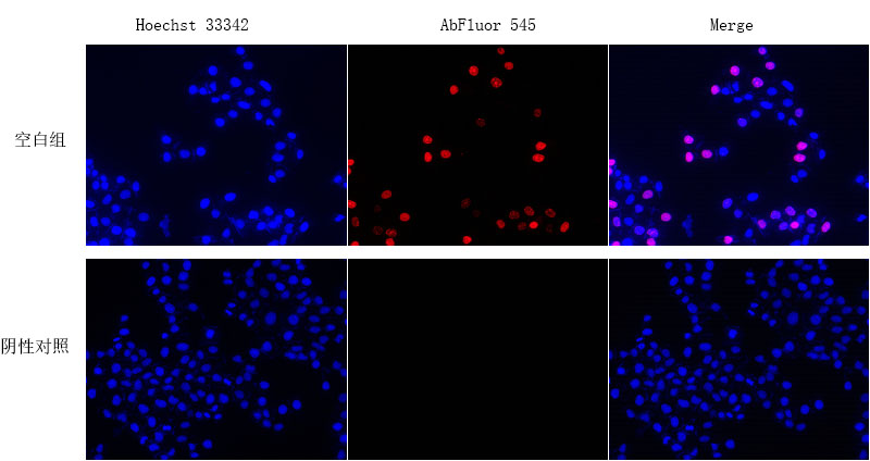
最準確的增殖檢測方法( proliferation-detection methods)是基於新合成 DNA 中核苷類似物(nucleoside analogues)的摻入和測量,其中溴脫氧尿苷 (bromodeoxyuridine, BrdU) 是常用的類似物。以嚴苛方法(HCl、熱或酵素)使 DNA 變性以暴露 BrdU 分子後,使用抗 BrdU 抗體對 BrdU 標記的 DNA 進行定量。此步驟非常耗時且難以一致地執行。嚴厲的處理也會對樣本的完整性和品質產生不利影響,這使得與其他抗體共染色具有挑戰性。細胞增殖EdU Image試劑盒(Cell Proliferation EdU Image Kit, 貨號KTA2031)( (Orange Fluorescence))是BrdU分析的一種新型替代方法。 EdU(5-乙炔基-2′-脫氧尿苷, EdU (5-ethynyl-2′-deoxyuridine) )是胸苷的核苷類似物,在活性DNA合成過程中被摻入DNA中。與BrdU分析相比,EdU-Click分析不是基於抗體的,因此不需要DNA變性(denaturation)(通常使用HCl或加熱或用DNase消化)來檢測摻入的核苷。細胞增殖EdU Image試劑盒不僅易於使用,而且更準確檢測基於click reaction,疊氮化物和炔烴之間的銅催(copper-catalyzed)化共價反應在30分鐘內完成。可用於顯微鏡成像、流式細胞儀和高通量篩選。

(文獻來源) TUC338 knockdown inhibits DLBCL cell proliferation. (a) Establishment of two DLBCL cell lines stably knocking down of TUC338. (b–g) Soft agar colony, EdU, and CCK-8 assays detecting colony formation, DNA synthesis, and cell viability, respectively. Scale bar = 50 μM, ∗∗P < 0.01, ∗∗∗P < 0.001.

(文獻來源) GLUT12 overexpression promotes GC proliferation and attenuates responses to everolimus. A The WB analysis to show GLUT12 expression of SGC-7901 and HGC-27 cells that were infected with lentivirus carrying vectors or GLUT12 overexpression plasmids. B-D The CCK-8 (B), colony formation (C) and EdU (D) assays to measure proliferation of cells as in (A).

(文獻來源) Effects of low dose GO on the proliferation of PC3 cells in vitro. (a) Representative images of the cell proliferation assay for PC3 cells under GO nanosheet (GO-L, GO-M, and GO-S) treatment at 0, 5, 10, 15, 20, and 50 μg/mL for 24 h with EdU doped. (b) Statistical data of the EdU positive ratios, n = 5. Scale bar = 500 μm. (c) Western blotting of expression levels of the PI3K/AKT/mTOR signal pathway-related proteins in PC3 cells post GO nanosheet (GO-L, GO-M, and GO-S) treatment at 0, 2, 5, 10, 15, 20, and 50 μg/mL for 24 h *: P < 0.05, **: P < 0.001, relative to untreated control.

(文獻來源) Sitagliptin inhibits proliferation and induces apoptosis in immortalized glioma cells. (A-D) EdU and Colony formation assays were performed to evaluate the effect of gradient concentrations of Sitagliptin on cell proliferation in T98G and U251 cells. Representative EdU and colony images are shown in A and C, respectively. Quantitative analyses of the percentage of EdU-positive cells and the number of colonies are shown in B and D, respectively. (E-H) Flow cytometry and TUNEL assays were used to analyze the effect of gradient concentrations of Sitagliptin on cell apoptosis in T98G and U251 cells. Representative flow cytometry histograms and TUNEL images are shown in E and G, respectively. Quantitative analyses of the percentage of apoptotic cells and the percentage of TUNEL-positive cells are shown in F and H, respectively. Scale bars: 200 µm. **P < 0.01, ***P < 0.001.

(文獻來源) Knockdown of CKAP2L expression inhibits KIRC cell proliferation (scale bar: 500 µm. *p < 0.05; **p < 0.01; ***p < 0.001; ****p < 0.0001). (A,B) Protein blotting images and quantitative data showed that the expression of CKAP2L in A498 and 786-O cells was significantly reduced after siRNA transfection. The uncropped original image can be obtained in Supplementary Fig. S7. (C) CCK8 assay detects the change in the proliferation ability of A498 and 786-O cells after CKAP2L knockdown. (D,E) Clone formation assay examined the change in the proliferation ability of A498 and 786-O cells after CKAP2L knockdown. (F,G) EdU incorporation assays assess the change in the proliferation ability of A498 and 786-O cells after CKAP2L knockdown.
![]()
Specification
| Product name | EdU Cell Proliferation Image Kit (Orange Fluorescence) |
| Applications notes | Cell Proliferation EdU Image Kit (Orange Fluorescence) is a novel alternative to the BrdU assay. EdU (5-ethynyl-2´-deoxyuridine) is a nucleoside analog of thymidine and is incorporated into DNA during active DNA synthesis. Comparing to BrdU assays, the EdU-Click Assays are not antibody based and therefore do not require DNA denaturation (typically using HCl or heat or digestion with DNase) for detection of the incorporated nucleoside. Detection is based on a click reaction, a copper-catalyzed covalent reaction between an azide and an alkyne, is complete within 30 minutes. |
Product Properties
| Kit components | • EdU (10mM) •BSA Wash Solution (5×) •AbFluor 545 azide •Reaction buffer (10×) •Copper Reagent •Reducing Agent •Hoechst 33342 (1000×) •Hydroxyurea |
| Features & Benefits | • Optimized negative control is provided to rule out false positives •No antibody needed. • No denaturation steps, preservation of cell morphology and DNA integrity. • Simple, reliable and time-saving than traditional methods. • Proprietary AbFluor 545 azide (Ex/Em = 546/565 nm)-good photostability and minimizing fluorescence quenching. • Optimized for fluorescent microscopy. |
| Storage instructions | Stable for at least 12 months at recommended temperature from date of shipment. Gel pack with blue ice. |
| Shipping | Gel pack with blue ice. |
| Precautions | The product listed herein is for research use only and is not intended for use in human or clinical diagnosis. Suggested applications of our products are not recommendations to use our products in violation of any patent or as a license. We cannot be responsible for patent infringements or other violations that may occur with the use of this product. |
Additional Information
| Background | The detection of cell proliferation is of utmost importance for assessing cell health, determining genotoxicity or evaluating anticancer drugs. Until now, measuring DNA synthesis directly is most accurate method of doing it, normally performed by incorporation of the nucleoside analog like [3H] thymidine or 5-bromo-2’-deoxyuridine to cells during replication, and then detected or visualized by autoradiography or with an anti-BrdU-antibody respectively. |
| 貨號 | 產品名稱 | 胞器染色位置 | 螢光波段 | |
| 貨號KTC4003 | TraKine™粒線體染色 | 粒線體(mitochondria) | Ex / Em = 490/523 nm | 增殖(proliferating)和非增殖細胞(non-proliferating cells) |
| 貨號KTC4100 | Pro活細胞微管蛋白染色 | 微管蛋白(Tubulin) | Ex/Em = 500/520 nm | 活細胞或固定之細胞 |
| 貨號KTC4001 | Abbkine 細胞膜染色 | 細胞膜 (plasma membrane) | Ex/Em = 484/501 nm | 活細胞、固定懸浮、貼附細胞 |
| 貨號BMD00063 | DAPI Staining Solution | 細胞核 dsDNA | Ex/Em: 358/461 nm (with DNA) | 染色活細胞和固定細胞 |
| 貨號KTC4210 | 活細胞Lysosome染色 | Lysosome | deep red fluorescent dye , Ex/Em:650/665nm | 染色活細胞和固定細胞 |