【ScyTek免疫染色劑】Histology products list
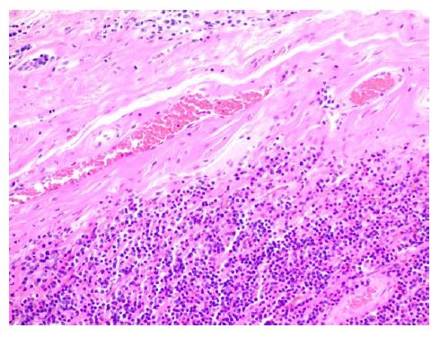
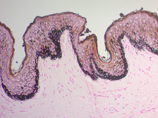
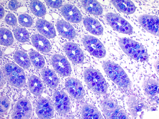
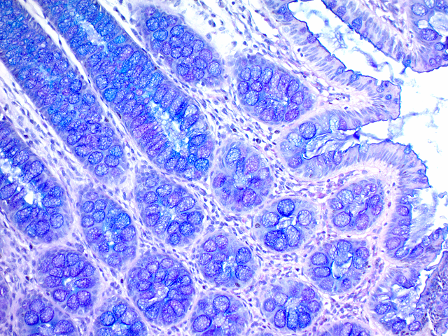
組織學常用組織化學染色 (Histochemistry stain) 使不同組織呈現不同顏色。組織化學染色溶液系列產品,提供不同組織之染色方法,例如蘇木紫-伊紅染色(Hematoxylin & Eosin stain),為最廣泛使用於組織病理之組織化學染色。Masson’s 染色,主要用於區別膠原纖維與其他組織。AB-PAS染色用於分類 gastric mucosal胃粘膜。Triphenyl tetrazolium chloride (TTC)氯化三苯基四氮唑(TTC)是一種脂溶性光敏化合物fat soluble light sensitive compound,,脫氫酶反應中dehydrogenase reaction的正常組織 normal tissue是紅色的,並且組織缺血 tissue ischemia是蒼白pale的。傳統組織化學染色試劑保存容易且作法簡單。可應用於石蠟切片Paraffin section; Section type、冷凍切片Frozen section細胞。
![]()
![]()
ScyTek Laboratories成立於1991年,其目標是成為生產生命科學試劑的世界級製造公司。從早期生產ELISA試劑開始,ScyTek逐漸擴大產品線,包括IHC試劑,特殊染色,緩沖液,Mounting Media,固定液,稀釋液和各種客製化試劑等等。從一開始,ScyTek就採用了嚴格的原料篩選流程,以確保所有製造的產品都是最高品質。2011年,ScyTek實驗室獲得了ISO 9001-2008認證,在2016年獲得了ISO 13485認證。
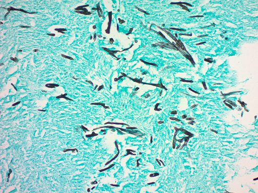
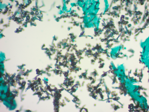
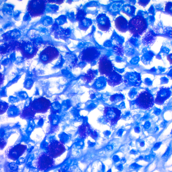
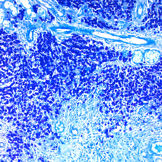
GMS 染色試劑盒 | Modified Gomori Methenamine-Silver Nitrate Stain貨號KAA-1 (125ML)
 Modified Gomori Methenamine-Silver Nitrate Stain(GMS 染色試劑盒)旨在用於真菌(fungi)、基底膜(basement membrane)和一些機會性生物(如卡氏肺囊蟲, Pneumocystis carinii)的組織學可視化。卡氏肺囊蟲是一種機會性病原體,可導致嚴重的肺病人類、狗、大鼠、小鼠和其他脊椎動物的疾病,具有獲得性、誘發性或遺傳性免疫缺陷綜合徵。此外,此程序將演示放線菌和相關種、諾卡氏菌小行星和某些包囊細菌。

![]() Procedure (Standard):
Procedure (Standard):
- Deparaffinize sections if necessary and hydrate to distilled water.
- Incubate slide in Chromic Acid Solution for 10 minutes.
- Rinse in tap water followed by 2 changes of distilled water.
- Incubate slide in Sodium Bisulfite Solution for 1 minute (to remove any residual chromic acid).
- Rinse in tap water followed by 2 changes of distilled water.
- Combine the following for a working GMS solution: 25 ml Silver Nitrate Solution (0.2%) 25 ml Methenamine Solution 2 ml Borax Solution Note: Mixed solution may not be stored for reuse later.
- Place working GMS solution in 60° centigrade water bath and allow temperature to equilibrate.
- Incubate slide in working GMS solution for 10-15 minutes. Using plastic forceps, dip slide in distilled water and check under a microscope for evaluation of silver impregnation. Fungi should be dark brown. If color is not sufficient, return the slide to working GMS solution for 2-3 minutes and check again.
- Rinse in 4 changes of distilled water.
- Incubate slide in Gold Chloride Solution for 15-30 seconds.
- Rinse in 4 changes of distilled water.
- Incubate slide in Sodium Thiosulfate Solution (5%) for 2 minutes.
- Rinse in tap water followed by 2 changes of distilled water.
- Incubate slide in Light Green Solution for 2 minutes.
- Rinse slide using absolute alcohol.
- Dehydrate in 2 changes of absolute alcohol, clear, and mount in synthetic resin. Note: Increasing incubation time and temperature of the Chromic Acid Solution reduces background silver staining. We suggest a 10min incubation at 60°C if there is unwanted background staining.
![]() Procedure (Microwave):
Procedure (Microwave):
Note: These instructions were developed using a standard 500 watt microwave oven. Heating times should be modified as needed depending on the microwave oven used. 1. Deparaffinize sections if necessary and hydrate to distilled water.
- Place slide in plastic coplin jar filled with Chromic Acid solution. Cap jar loosely!
- Place jar in microwave oven and heat on high power for 10 seconds. Allow slide to remain in warm solution for 3 minutes.
- Rinse in tap water followed by 2 changes of distilled water.
- Incubate slide in Sodium Bisulfite solution for 1 minute (to remove any residual chromic acid).
- Rinse in tap water followed by 2 changes of distilled water.
- Combine the following for a working GMS solution: 25 ml Silver Nitrate 25 ml Methenamine 2 ml Borax Solution Note: Mixed solution may not be stored for reuse later.
- Place working GMS solution (loosely capped) in microwave oven for 40 seconds. Remove and pour several times between coplin jar and a clear graduated cylinder to mix thoroughly (use protective glove to avoid burning hand). Mixed solution remains in coplin jar.
- Incubate slide in working GMS solution (heated) for 2-6 minutes until the tissue is medium brown in color. Using plastic forceps, dip slide in distilled water and check under a microscope for evaluation of silver impregnation. Fungi should be dark brown. If color is not sufficient, return the slide to working GMS solution for 1-2 minutes and check again. Reheat solution if needed.
- Rinse in 4 changes of distilled water.
- Incubate slide in Gold Chloride solution for 15-30 seconds.
- Rinse in 4 changes of distilled water.
- Incubate slide in Sodium Thiosulfate for 2 minutes.
- Rinse in tap water followed by 2 changes of distilled water.
- Incubate slide in Light Green Solution for 2 minutes.
- Rinse slide using absolute alcohol.
![]()
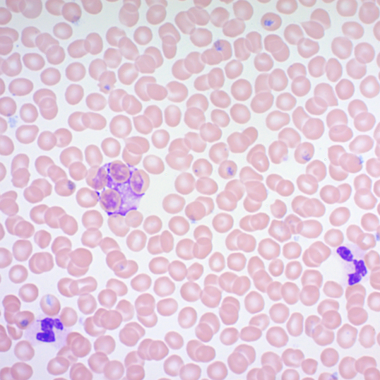
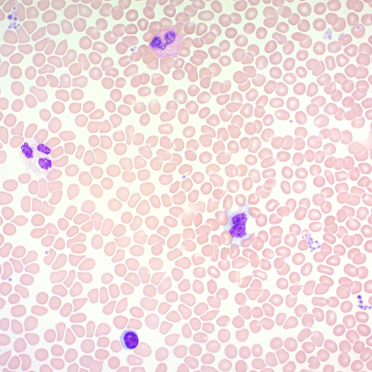
Wright-Giemsa Stain Kit試劑盒 | 貨號WGK-1 (500ml) WGF-2 (30ML)
WGS500 Wright-Giemsa Solution 500 ml
PBM500 Phosphate Buffer Solution (pH 6.8) 500 ml (x2)
Wright-Giemsa Stain Kit 操作手冊下載
產品介紹: Wright-Giemsa Stain Kit 旨在用於血塗片(blood smears)、骨髓(bone marrow)和血液寄生蟲(blood parasites)的差異染色。
紅血球(Erythrocytes):粉紅棕褐色
白血球(Leukocytes):藍紫色
中性粒細胞(Neutrophils):細胞質(cytoplasm)中呈淡紫色顆粒。
嗜酸性粒細胞(Basophils):細胞質(cytoplasm)中亮紅-紅-橙色顆粒。
嗜鹼性粒細胞(Basophils):細胞質(cytoplasm)中的深紫色顆粒。
血小板(Platelets):淡藍色細胞質(cytoplasm)中的紫紫色顆粒。Wright-Giemsa Stain Kit is intended to be used for differential staining of blood smears, bone marrow and blood parasites.
| Applications | • Histological applications • For in vitro diagnostic use |
| Features & Benefits | For differential staining of blood smears, bone marrow and blood parasites. |
| Kit Components | • Wright-Giemsa Solution • Phosphate Buffer Solution (pH 6.8) |
| Storage Conditions | RT |
| Shipping Conditions | RT |
| USAGE | For Research Use Only! Not For Use in Humans. |
Procedure(標準):
Prepare Working Wright-Giemsa Solution by mixing 25ml of Wright-Giemsa Solution (cat# WGS) with 25ml of Phosphate Buffer Solution, pH 6.8 (cat# PBM).
- 在乾淨的顯微鏡載玻片(microscope slide)上塗抹一小滴血,然後風乾。
- 將其置於無水甲醇(Methanol)中 5 分鐘進行固定。
- 將載玻片放入染色盤中並用 Working Wright-Giemsa Sol 浸漬 5 分鐘。注意:偶爾攪拌載玻片以確保正確染色。
- 在去離子/蒸餾水中沖洗載玻片。
- 用 pH 6.8 的磷酸鹽緩衝溶液(Phosphate Buffer Solution, pH 6.8)沖洗玻片,直到沒有污漬流走。
- 讓載玻片在 pH 6.8 的磷酸鹽緩衝溶液(Phosphate Buffer Solution, pH 6.8)中再停留 1 分鐘。
- 將載玻片浸入蒸餾水中並在室溫下風乾。
- 在二甲苯(Xylene)或二甲苯替代品(Xylene Substitute)中浸幾次玻片。
- 安裝在合成樹脂(synthetic resin)中。
穀胱甘肽過氧化物酶活性測定 | FRAP檢測 | 總膠原蛋白分析 | Dopamine多巴胺檢測 | 三酸甘油酯分析 | Cholesterol Efflux膽固醇排出螢光分析 | 脂質過氧化(MDA)測定 | 支鏈氨基酸 BCAA檢測
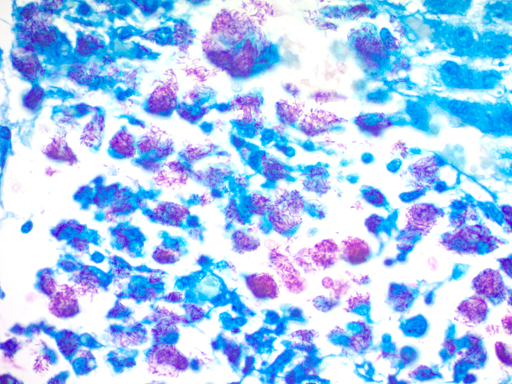
Gram Stain Kit (MODIFIED BROWN AND BRENN) | BBS-2
產品介紹: 革蘭氏染色(Gram Staining)用來鑒別細菌的一種方法,利用細菌細胞壁上的生物化學性質不同,分成革蘭氏陽性(Gram Positive)與革蘭氏陰性(Gram Negative)。用於鑑別革蘭氏陰性和革蘭氏陽性。Gram Stain Kit (MODIFIED BROWN AND BRENN) 應用原理: 細菌的陽性和陰性細胞壁均由肽聚醣(peptidoglycan)組成(革蘭氏陽性細胞壁較厚),並且兩者都將吸收結晶紫(crystal violet)。革蘭氏陰性菌在肽聚醣壁的外部有一層脂多醣(lipopolysaccharide),在丙酮沖洗液中被破壞,從而使結晶紫(crystal violet)得以區分。這使革蘭氏陰性細菌吸收鹼性品紅染色(basic fuchsin stain)。


革蘭氏陽性細菌:藍色
革蘭氏陰性菌:紅色
其他組織:淡黃色-粉紅色
核:紅色
流程可概括為:染色-脫色-複染。(標準操作請依據原廠網站)
1.如有必要,對切片進行脫蠟(Deparaffinize sections)處理,然後水合成蒸餾水。
2.用龍膽紫溶液(Gentian Violet Solution)覆蓋組織切片,孵育2分鐘。
3.用蒸餾水沖洗玻片,以去除多餘的stain。
4.用Lugol’s Iodine Solution的碘溶液覆蓋組織切片,並孵育1分鐘。
5.在流動的自來水中沖洗滑片,以除去過量的碘(Iodine)。
6.浸入蒸餾水中1次。
7.滴加Gram的脫色劑(Gram’s Decolorizer dropwise),直到顏色不再滲出為止。
8.在蒸餾水中快速沖洗載玻片(2-5秒)。
9.用番紅O(Safranin O Solution)溶液覆蓋組織切片並孵育4分鐘。
10.在蒸餾水中快速沖洗載玻片以去除多餘的stain。
11.浸入無水酒精一次並吸乾。
12.用Picric Acid – Acetone Solution(0.1%)覆蓋組織切片,並在攪拌下孵育2-10秒。
13.用3種無水酒精(absolute alcohol)迅速脫水。
14.清除2種二甲苯或二甲苯替代物的更換,然後裝入合成樹脂(synthetic resin)中。
亞甲基藍溶液(Methylene Blue Solution) | 貨號MBS125 125ml
Methylene Blue Solution
產品介紹: 優化之亞甲基藍溶液(Methylene Blue Solution),可在以下情況下獲得出色的結果;可用於各種程序,例如(Ziehl-Neelsen)、Fite和Gram染色。亦可用於染色各種細菌,如白喉棒狀桿菌(Corynebacterium diphtheria),流感嗜血桿菌(Haemophilus influenza),奈瑟氏菌(Neisseria)和白細胞(leukocytes)。 該試劑產生深藍色的核(nuclei),具有非常淺的藍色細胞質(cytoplasm)。
Nuclei: Dark Blue
Cytoplasm: Light Blue
Corynebacterium Diphtheria: Blue Granules
Escherichia Coli: Blue Rods
Streptococcus Pyogenes: Blue Cocci
避免與皮膚和眼睛接觸。吞食有害
甲苯胺藍 (Toluidine Blue Solution) | 貨號TQF125 (125ml)
產品介紹: 甲苯胺藍(Toluidine Blue Solution)是鹼性噻嗪異染色染料,對酸性組織成分具有高親和力。它使核酸藍色和多醣染成紫色,並且還增加組織學載玻片圖像的清晰度。甲苯胺藍溶液設計用於酸性物質(acidic substances)的變色染色。
染色結果顯示:酸性碳水化合物是變色的(粉紅色至紅色或紫色), 細胞核和細胞質是藍色的。結締組織粘蛋白,軟骨的磨碎物質,肥大細胞顆粒和許多上皮。粘蛋白是變色的。Acidic carbohydrates are metachromatic (pink to red or purple). Nuclei and cytoplasm are blue.Connective tissue mucins, ground substances of cartilage, mast cell granules and many epithelial mucins are metachromatic.
Procedure操作方法 (若有不同,以原廠操作手冊為標準)
- Deparaffinize sections if necessary and hydrate to distilled water.
- Incubate slide in Periodic Acid Solution for 10 minutes (Note: Do Not Reuse Periodic Acid Solution).
- Rinse thoroughly in distilled water.
- Incubate slide in Sodium Metabisulfite Solution for 5 minutes.
- Rinse thoroughly in distilled water.
- Incubate slide in Alcian Yellow Solution for 5-10 minutes.
- Rinse slide thoroughly in distilled water.
- Incubate slide in freshly prepared Toluidine Blue Working Solution for 3-5 minutes (Note: Do Not Reuse Toluidine Blue Working Solution).
- Rinse thoroughly in distilled water.
- Dehydrate quickly in 2 changes of absolute alcohol.
- Clear, and mount in synthetic resin.
Trichrome染色 | AFB)染色 | Alcian Blue – PAS染色 | Alcian Blue(pH 0.5)染色 | Amyloid Stain | Fite’s Stain Kit | Fontana-Masson染色
組織化學染色 (Histochemistry stain) – Eosin Y Solution (Modified Alcoholic) 貨號EYB500
產品介紹: Eosin Y Solution(Modified Alcoholic)適用於組織學和細胞學應用。 這種新配方的曙紅(Eosin)提供了傳統酒精配方( traditional alcoholic formula)的優點,並顯著改善了可用性。 優點包括較低的蒸發速率,較好的顏色圖案,減少溢出容器,手和檯面的傾向,以及保持在組織切片上的改善的表面張力。Eosin Y Solution (Modified Alcoholic) is intended for use in histology and cytology applications. This newly formulated Eosin provides the benefits of a traditional alcoholic formula with significant improvements in usability. Advantages include lower evaporation rate, better color patterns, reduced tendency to spill over container, hands, and countertops, and improved surface tension to remain on tissue section.
細胞質:淡粉色
膠原蛋白:粉紅色
肌肉:粉紅/玫瑰
紅細胞:粉紅色/紅色
Eosin Y Solution (Modified Alcoholic)程序:
- 必要時部分脫蠟 (Deparaffinize sections) 並與蒸餾水混合。
- 將載玻片浸入無水酒精中並過量吸乾。
- 使用足夠的Eosin Y溶液(改良酒精)使組織切片完全覆蓋過量並孵育2-3分鐘。
4.用無水酒精沖洗載玻片。
5.在3次無水酒精變化中脫水。
- Clear slide並mount in synthetic resin。
組織化學染色 (Histochemistry stain) – Eosin Y Solution (Aqueous)-貨號EYQ500, 500ml
產品介紹: Eosin Y Solution (Aqueous)適用於細胞質的組織學 (histological demonstration),通常用作蘇木精(Hematoxylin)的複染劑。 如果使用得當,可以獲得各種粉紅色,以幫助組織成分的可視化。紅血球細胞(Erythrocytes),膠原蛋白(collagen)和肌肉或上皮細胞的細胞質(cytoplasm)會染上不同深淺的粉紅色。
細胞質(Cytoplasm):粉紅色至紅色
紅細胞(Erythrocytes):粉紅色到紅色
細胞核(Nuclei):黑/藍(蘇木精)
Eosin Y Solution(Aqueous)程序:
1.必要時將部分脫蠟(Deparaffinize sections)並與蒸餾水(distilled water)混合。
2.如果切片是Zenker固定的,用碘去除氯化汞晶體(chloride crystals)並用硫代硫酸鈉(sodium thiosulfate)清除。用流動的自來水沖洗。
3.用蒸餾水沖洗載玻片。
5.在蘇木精,Mayer’s(HMM125)中染色5分鐘。
6.用流動的水沖洗載玻片2-3分鐘。
7.塗抹藍光試劑(BRT125)30秒。
8.用蒸餾水沖洗。
9.在Eosin Y溶液中染色載玻片5分鐘。
10.用95%酒精快速沖洗,然後用無水酒精沖洗2分鐘。
11.清除,並安裝在合成樹脂(synthetic resin)中。
組織化學染色 (Histochemistry stain) – Eosin Y Solution (Alcoholic)-貨號EYA500, 500ml
產品介紹: Eosin Y Solution (Alcoholic)適用於細胞質的組織學 (histological demonstration),通常用作蘇木精(Hematoxylin)的複染劑。 如果使用得當,可以獲得各種粉紅色,以幫助組織成分的可視化。紅血球細胞(Erythrocytes),膠原蛋白(collagen)和肌肉或上皮細胞的細胞質(cytoplasm)會染上不同深淺的粉紅色。
Cytoplasm: Pink to Red
Erythrocytes: Pink to Red
Nuclei: Black/Blue (Hematoxylin)
細胞質:粉紅色至紅色
紅細胞:粉紅色到紅色
細胞核:黑/藍(蘇木精)
Eosin Y Solution程序:
1.在優選的蘇木精(Hematoxylin)中染色1-5分鐘
注意:通常較長的孵育時間會產生較深的染色。 (ScyTek Cat#HMM)
2.用自來水或去離子水(deionized water)沖洗載玻片。
3.如果需要,藍色試劑藍色15-30秒(ScyTek Cat#BRT)
組織化學染色 (Histochemistry stain) -Fast Green Solution (貨號FGN125, 125ml)
產品介紹: Fast Green Solution (貨號FGN125),為染色膠原蛋白(collagen)和網狀纖維(reticular fibers)。 使用經認證的Fast Green FCF製造,提供single component即用型試劑(Ready-To-Use reagent)。
Fast Green Solution程序:
1.必要時對組織進行脫蠟,並與蒸餾水混合。
2.根據所需的染色強度將組織或細胞染色1-5分鐘。
3.如果需要,在1%乙酸溶液中分化。 (ScyTek Cat#AAE)
4.在去離子水中短暫沖洗並脫水至無水乙醇。
5.將滑塊固定並並以封片劑 (permanent mounting medium)封片。
組織化學染色 (Histochemistry stain) -Safranin O Solution 貨號SOH500, 500ml

產品介紹: safranin番紅,也稱作番紅O或基本紅2)是個用在組織學和細胞學的生物染色劑。番紅Safranin O Solution通常用於複染核紅(Hucker’s Counterstain)。將所有的細胞核染成紅色。
Safranin O Solution程序:
1. Re-hydrate 組織切片。
2.番紅O (Safranin O) 溶液組織切片染色5分鐘。
3.95%酒精(95% alcohol)沖洗1分鐘,然後用無水乙醇(absolute alcohol)沖洗1分。
4.用聚苯乙烯(xlylene)清除,並以封片劑(permanent mounting medium)封片。
Hematoxylin and Eosin Stain Kit蘇木精和伊紅染色試劑盒 | 貨號HAE-1-IFU
產品描述:蘇木精和伊紅染色試劑盒(Hematoxylin and Eosin Stain Kit)旨在用於組織學(histology)和細胞學(cytology)應用。該試劑盒中包含一種新配方的曙紅(Eosin),可提供傳統酒精配方 (traditional alcoholic formula) 的優點,並顯著提高可用性。蘇木精產生清脆,強烈的藍色核,與曙紅染色的細胞質形成最佳對比。Advantages include lower evaporation rate, better color patterns, reduced tendency to spill over container, hands, and countertops, and improved surface tension to remain on tissue section. Our Hematoxylin produces crisp, intense blue nuclei providing optimal contrast to the Eosin stained cytoplasm. Cytoplasm: Light Pink
Collagen: Pink
Muscle: Pink/Rose
Erythrocytes: Pink/Red
Nuclei: Blue
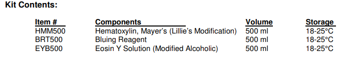
Mayer’s Hematoxylin (Lillie’s Modification)
產品描述: Mayer’s Hematoxylin (Lillie’s Modification) is a progressive nuclear hematoxylin stain with several histological applications. Nuclei should staining is strong, clean, crisp, and no differentiation is needed. Bluing reagent (Catalog: BRT) may be used following hematoxylin to “blue” or alter the shade of hematoxylin from a purple to blue
Nuclei: Purple
Nuclei after Bluing: Blue
Nuclei after Eosin: Blue to Violet
Fontana-Masson染色 | 貨號FMS-1 (Kit 125ml) /貨號FMS-2 (Kit 30ml)
產品介紹: Fontana-Masson染色試劑盒適用於paraffin和冷凍切片中( frozen sections) 的 Argentaffin cells和黑色素 ( Melanin) 的組織學可視化。

Argentaffin Cells: Black
Melanin: Black
Nuclei: Red
Cytoplasm: Light Pink
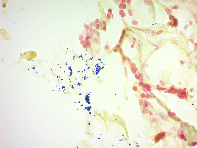
Fite’s Stain Kit染色試劑盒 | 貨號FLS-1 (125ml) / FLS-500 (500ML)
產品介紹: Fite的染色試劑盒(用於Leprosy麻風病)旨在用於麻風分枝桿菌( mycobacterium leprae)(leprosy麻風病)和諾卡氏菌(Nocardia)的組織學可視化。
Lepra bacillus: Red
Nocardia: Red
Background: Blue
Amyloid Stain Kit澱粉樣染色 | 貨號AMY-2 (Kit) (以下規格為kit 30ML包裝/貨號AMY-2)
產品介紹: 澱粉樣染色試劑盒(Congo Red , 剛果紅)旨在用於組織切片中澱粉樣蛋白 (amyloid )的組織學可視化。 在偏光顯微鏡 (polarizing microscope)下檢查導致澱粉樣蛋白(amyloid)的綠色雙折射 (green birefringence )。澱粉樣蛋白:紅色到粉紅色。紅細胞:淡橙色。嗜酸性粒細胞顆粒:橙色至紅色。細胞核:藍色。
Amyloid: Red to Pink
Erythrocytes: Light Orange
Eosinophil Granules: Orange to Red
Nuclei: Blue

Alcian Blue(pH 0.5)染色試劑盒 | 貨號AFS-1 (Kit)
產品介紹:Alcian Blue(pH 0.5)染色試劑盒旨在用於非常強硫酸化的粘膜物質 ( sulfated mucosubstances) 的組織學可視化。非常強烈硫酸化的物質:藍色。細胞核:紅色。背景:粉紅色

Very Strongly Sulfated Mucosubstances: Blue
Nuclei: Red
Background: Pink
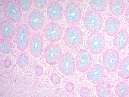
Alcian Blue – PAS染色試劑盒 | 貨號APS-2 (100slide) APS-1 (Kit) 以下規格內容為貨號APS-1 (kit)
產品介紹: Alcian Blue – PAS染色試劑盒適用於硫酸化(sulfated )和羧化 (carboxylated) 酸性粘多醣 (mucopolysaccharides),硫酸化(sulfated )和羧化唾液粘蛋白(carboxylated sialomucins)(glycoproteins, 糖蛋白)和中性粘蛋白(neutral mucins)的組織學可視化。酸性硫酸化物質:藍色。透明質酸:藍色。Sialomucins:藍色。中性粘蛋白:洋紅色。酸性和中性粘蛋白的混合物:藍色 – 淡紫色取決於主導實體。

Acidic Sulfated Mucosubstances: Blue
Hyaluronic Acid: Blue
Sialomucins: Blue
Neutral Mucins: Magenta
Mixtures of Acidic and Neutral Mucins: Blue – Mauve depending on dominant entity.
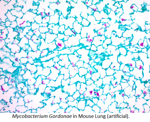
酸性快速細菌(AFB)染色試劑盒 | 酸性快速細菌/結核桿菌染色 貨號FAB-1/FAB-2/FAB-500 (以下規格為30ML包裝/貨號FAB-2)
產品名稱: 酸性快速細菌(Acid Fast Bacteria, AFB)染色試劑盒旨在用於酸性快速細菌 ( Acid Fast Bacteria) 和結核桿菌 (Tubercle Bacilli) 的組織學可視化。 快速的15分鐘程序。 耐酸生物的類脂質膠囊 ( lipoid capsule) takes up carbol fuchsin並抵抗脫色。
Acid Fast Organisms: Bright Red
Background: Light Green

石蠟切片Paraffin section; Section type、冷凍切片Frozen section細胞
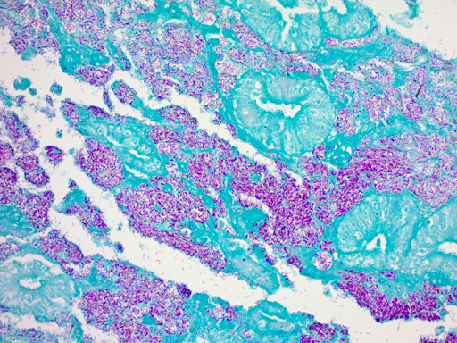
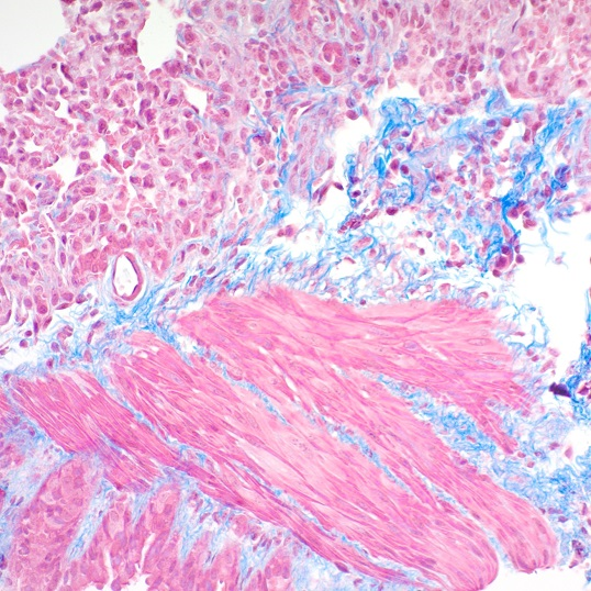
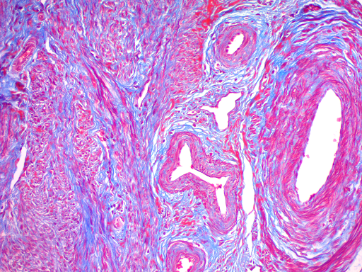
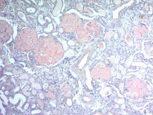
Trichrome染色試劑盒(改良Masson’s)染結締組織 | 貨號TRM-2/TRM-1/TRM-500
產品名稱: Trichrome染色試劑盒(改良Masson’s)適用於組織學實驗室,用於組織切片中膠原結締組織纖維的染色和隨後的可視化。 這種Trichrome Special Stain試劑盒可用於福爾馬林固定,石蠟包埋或冷凍切片。傳統Masson染色法是利用兩種或三種陰離子染料混合一起或先後作用對組織進行染色的一種方法,也是膠原纖維權威而經典的技術方法。 Trichrome染色試劑盒(改良Masson’s)產品應用: 染色主要用於膠原纖維collagen fibers 和肌纖維muscle fibers的差異染色。 膠原和軟骨 Collagen and cartilage呈藍色,肌纖維 cellulose ,纖維素 cellulose 和紅細胞 red blood cell 染成紅色,細胞核nuclei 染成藍黑色。
Collagen: Blue
Muscle Fibers: Red
Nuclei: Black/Blue
石蠟切片Paraffin section; Section type、冷凍切片Frozen section細胞