【細胞衰老檢測】Cell Senescence Kit (β-Galactosidase Assay) 貨號KTA3010
細胞衰老檢測試劑盒(β-半乳糖苷酶) | 貨號KTA3010
Cell Senescence Kit (β-Galactosidase Assay)
細胞衰老(Cellular senescence)是正常細胞停止分裂(cell divide)的現象。衰老的細胞(senescent cells)不再能夠複製(replicate),但它們仍保持代謝活性(metabolically active),但因阻滯於G1期,失去了對有絲分裂原的反應能力和合成DNA的能力,不能進入S期。並且與衰老相關的β-半乳糖苷酶活性染色(senescence-associated beta-galactosidase activity)呈陽性,這被認為是細胞衰老的生物標記(biomarker)。細胞的衰老(Cellular senescence)可以通過激活癌基因(oncogenes)和細胞-細胞融合(cell-cell fusion)來實現,DNA對活性氧(reactive oxygen species , ROS)升高的反應會導致DNA損傷(DNA damage),而不是僅僅依賴於細胞分裂的數目。在正常衰老過程(normal aging)中,組織中的衰老細胞數量顯著增加。
產品描述: Abbkine細胞衰老檢測試劑盒(β-半乳糖苷酶, β-Galactosidase)提供了基於衰老過程中pH值為6的衰老相關β-半乳糖苷酶(SA-β-Gal)活性上調的試劑,用於檢測衰老細胞(senescent cells)或組織(tissues)。細胞或組織的老化可以在普通的光學顯微鏡下觀察。 SA-β-Gal僅存在於衰老細胞(senescent cells)中,在衰老前,靜止,腫瘤或永生細胞中無法檢測到。

Cellular senescence is a phenomenon in which normal cells cease to divide. The senescent cells can no longer replicate, but they still maintain metabolically active and stain positive for senescence-associated beta-galactosidase activity, which is considered to be a biomarker of cellular senescence. Cellular senescence can be achieved by the activation of oncogenes and cell-cell fusion, DNA damage in response to elevated reactive oxygen species (ROS), rather than relying solely on the number of cellular divisions. During normal aging, the number of senescent cells in tissues increases significantly. This kit provides reagents for detecting the senescent cells or tissues based on the up-regulation of senescence-associated β-galactosidase (SA-β-Gal) activity at pH 6 during aging. The aging of cells or tissues can be observed under a common optical microscope. SA-β-Gal is present only in senescent cells and is undetectable in pre-senescent, quiescent, tumor or immortal cells.
商品需用儀器測量 酵素免疫分析儀請按此 (標配405、450、492、630 nm, 可擴增至7個濾片)

活細胞和死細胞雙染色套組 | CCK-8細胞增殖和細胞毒性試劑盒 | LDH細胞毒性測定試劑盒 | 細胞衰老檢測試劑盒(β-半乳糖苷酶 | 細胞增殖EdU Image試劑盒
 活細胞和死細胞雙染色套組 | Live and Dead Cell Double Staining Kit貨號KTA1001
Detect using either microscopy or FACS.
活細胞和死細胞數量的定量是細胞生物學研究中不可缺少的工具。死細胞對於研究生長控制和細胞死亡非常重要。Abbkine 的活/死細胞可行性測定試劑盒 (KTA1001)提供了一種雙色螢光方法,該方法基於使用兩種不同的染料同時測定活細胞和死細胞。
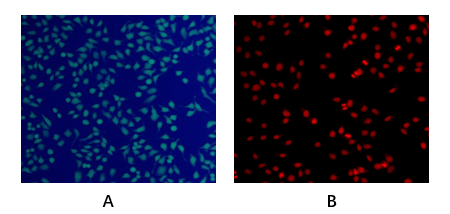
產品描述:Abbkine 活細胞和死細胞雙染色試劑盒 (Live and Dead Cell Double Staining Kit, KTA1001) 提供了一個方便的測定評估死活細胞的快速方法,基於同時測定活細胞和死細胞與兩種Probe,測量細胞健康、血漿膜完整性、和細胞內酯酶活性。該試劑盒利用LiveDye,一種細胞滲透的綠色螢光染料(Ex/Em = 488/530 nm),染色活細胞和核染料,一種細胞不可滲透的紅色螢光染料(Ex/Em = 535/617),以染色死細胞。
引用文獻
1. Ocular surface repair using decellularized porcine conjunctiva
L Zhao, Y Jia, C Zhao. Acta biomaterialia, 2019 – Elsevier
2. Grafting antibacterial polymer brushes from titanium surface via polydopamine chemistry and activators regenerated by electron transfer ATRP
Shuangshuang Wang, Jixuan Song, Yuchao Li, et al. Reactive and Functional Polymers. July 2019, 140: 48-55.
 CCK-8細胞增殖和細胞毒性試劑盒 | 貨號 KTC011001
CCK-8; WST-8; CCK8; WST8
產品描述:Abbkine Cell Counting Kit-8(CCK-8)設計用於檢測細胞增殖和細胞毒性。細胞計數試劑盒-8(CCK-8)使用高度水溶性的四唑鹽WST-8:[2-(2-甲氧基-4-硝基苯基)-3-(4-硝基苯基)-5-(2,4-二磺基苯基 )-2H-四唑鎓單鈉鹽]通過還原轉化為水溶性橙色甲dye染料。 CCK-8具有低毒性,並且允許用於測定細胞增殖和細胞毒性測定中活細胞數量的靈敏比色測定。 細胞中脫氫酶產生的甲dye染料的量與活細胞的數量成正比。

- Formulation
Liquid solution
- Kit components
- CCK-8 solution
- Features & Benefits
- One-bottle, ready-to-use solution.
• The product (formazan) produced by WST-8 is water soluble, No organic solvents or isotopes required. And the formazan is stable and safe.
• The detection sensitivity using CCK-8 is higher than assays using other tetrazolium salts such as MTT, XTT, MTS or WST-1.
• The same cells can be used for other cell assays because of the low toxicity of CCK-8. - Storage instructions
CCK-8 is stable over one year at 0-5°C with protection from light. Store it at -20°C for longer storage. Repeated thawing and freezing causes an increase in the background, which interferes with the assay. Please store the kit at 0-5°C for frequent use.
- Shipping
Gel pack with blue ice.
注意事項
- 小心不要將氣泡引入Well,因為它們會干擾O.D. 讀。
- 孵育時間因細胞的類型和數量而異。 通常,白細胞的著色較弱,因此可能需要較長的培養時間(長達4小時)或大量細胞(〜105細胞/孔)。
- 如果培養基的顏色或PH由於長期培養而改變,請在添加CCK-8時更換培養基。
LDH Cytotoxicity Assay Kit
乳酸脫氫酶(LDH)是一種氧化還原酶,可催化相互轉化丙酮酸和乳酸的結合,同時伴隨著NADH和NAD +的相互轉化。有5個LDH,LDH1至LDH5的不同同工酶,每個同工酶由4個亞基組成,可能是2個不同類型的M和H亞基。這些亞基由兩個不同的基因編碼。M亞基由基因LDHA編碼,而H亞基由LDHB編碼。通常,LDH2是血清中的主要形式。LDH為Plasma membrane損傷的定量方式,用來評估細胞死亡(Cell death)或細胞毒性(cytotoxicity)。 LDH(Lactate dehydrogenase, LDH)是一種穩定的酶,存在於所有細胞類型中,並在lasma membrane受損後迅速釋放到細胞培養基中。因此,LDH是細胞毒性研究中使用最廣泛的標記。
產品描述:Abbkine LDH細胞毒性測定試劑盒 (Abbkine LDH Cytotoxicity Assay Kit, KTA1030)為研究細胞毒性提供了一種簡便的比色測定法。該測定基於在NADH偶聯的酶促反應中四唑鹽MTT的還原成MTT的還原形式,該MTT在565nm處表現出最大吸收。產生的顏色的強度與裂解的細胞數直接相關。在典型的細胞毒性測定中,將靶細胞與細胞毒性化學試劑或細胞毒性細胞(NK細胞,細胞毒性T細胞)一起培養,以誘導靶細胞死亡和LDH釋放。將含有LDH的上清液轉移至新的96孔測定板的孔中,並與LDH反應溶液混合。在室溫下孵育30分鐘後,使用酶標儀讀取565nm(A565)的吸光度。
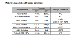
- Kit components• Assay Buffer
• Lactic Acid Solution
• MTT Solution
• PES Solution
• LDH Positive Control
• NAD+ Solution
• Triton X-100 (10%) - Features & Benefits• Measures cell death in response to chemical compounds or environmental factors.
• LDH release from the cytosol into the medium catalyzes a color-forming reaction.
• Detect lysis of 10,00-100,0000 cells per well. - Usage notesPerforming an initial titration experiment to determine the optimal number of cells per well of the target cell you plan to use.
- Storage instructionsStore at -20°C and Keep in dark. The Kit has a storage time of 6 months from receipt
細胞增殖EdU Image試劑盒 | Cell Proliferation EdU Image Kit
【DNA合成/細胞增殖檢測】 EdU Image試劑盒-BrdU分析新型替代方法
細胞增殖(cell proliferation)的檢測,對於評估細胞健康(cell health),確定遺傳毒性(genotoxicity)或評估抗癌藥物(anticancer drugs)至關重要。到目前為止,直接測量DNA合成(DNA synthesis)是最準確的方法,通常是通過在複製過程中將[3H]胸苷(thymidine)或5-溴-2′-脫氧尿苷(5-bromo-2’-deoxyuridine)等核苷類似物(nucleoside analog like)摻入細胞中,然後通過放射自顯影或分別使用抗BrdU抗體(anti-BrdU-antibody)。EdU(5-乙炔基-2′-脫氧尿苷)是BrdU(5-bromo-2′-脫氧尿苷)的新型替代品。它被取代胸苷摻入到新合成的增殖細胞DNA中。將5-EdU摻入到增殖細胞中,然後使用點擊連接用螢光疊氮化物進行檢測。與BrdU的使用相反,新方法不需要樣品固定或DNA變性。可用於研究中樞神經系統中的細胞增殖。

產品描述:
細胞增殖EdU Image試劑盒(Cell Proliferation EdU Image Kit, 貨號KTA2030)(綠色螢光)是BrdU分析的一種新型替代方法。 EdU(5-乙炔基-2′-脫氧尿苷)是胸苷的核苷類似物,在活性DNA合成過程中被摻入DNA中。與BrdU分析相比,EdU-Click分析不是基於抗體的,因此不需要DNA變性(denaturation)(通常使用HCl或加熱或用DNase消化)來檢測摻入的核苷。檢測基於click reaction,疊氮化物和炔烴之間的銅催(copper-catalyzed)化共價反應在30分鐘內完成。
- Kit components
- EdU (10mM)
• AbFluor 488 azide
• 10×Reaction buffer
• Copper
• Reducing Agent - Features & Benefits
- No antibody needed.
• No denaturation steps, preservation of cell morphology and DNA integrity.
• Simple, reliable and time-saving than traditional methods.
• Proprietary AbFluor 488 azide (Ex/Em = 501/525 nm)-good photostability and minimizing fluorescence quenching.
• Optimized for fluorescent microscopy. - Storage instructions
Stable for at least 12 months at recommended temperature from date of shipment. Gel pack with blue ice.
- Shipping
Gel pack with blue ice.
引用文獻
In vitro mitochondrial-targeted antioxidant peptide induces apoptosis in cancer cells. W Zhan, X Liao, L Li. Onco Targets Therv.12
Perilaldehyde activates AMP‐activated protein kinase to suppress the growth of gastric cancer via induction of autophagy
Y Zhang, S Liu, Q Feng. JOURNAL OF CELLULAR BIOCHEMISTRY