【凋亡細胞分析Annexin V&TUNEL】Apoptosis Assay Cocktail貨號 KTD102-EN
凋亡細胞分析(Annexin V法和TUNEL法) | Apoptosis Assay Cocktail貨號 KTD102-EN
凋亡細胞會產生許多重要的變化,包括細胞收縮(cell shrinkage)、細胞質凝集(cytoplasmic agglutination)、DNA斷裂(DNA disruption)和膜囊泡(membrane vesicles)的形成,並導致細胞成分分離形成凋亡小體(apoptotic dies),最終被巨噬細胞(macrophages)或實質細胞吞噬(parenchyma cells)Abbkine 基於新一代 AbFluor™ 螢光染色(AbFluor™ fluorescent staining)和高效重組蛋白技術(recombinant protein technology),開發了一種雞尾酒套裝,可同時檢測早期和晚期細胞凋亡的不同階段。 Vermes 是第一個使用 Annexin V 染色(Annexin V staining)特異性鑑定外翻細胞膜上的磷脂酰絲氨酸 (PS) 的研究人員,Annexin V 檢測被全球研究人員認為是最經典的方法。此外,DNA片段化(DNA fragmentation)是細胞凋亡晚期的一個重要特徵,基於DNA片段化檢測(DNA fragmentation detection)的TUNEL方法被認為是細胞凋亡研究的又一金標準。適用於組織(tissues)和細胞(cells)等不同樣品。高靈敏度(High sensitivity)、通用的細胞凋亡檢測試劑盒(universal apoptosis detection kit),可有效區分細胞凋亡(apoptosis)與壞死(necrosis)。同時,針對細胞凋亡檢測和結果的非標準化,我們開發了一種專利的細胞凋亡檢測陽性對照物質(positive control),並結合到細胞凋亡檢測雞尾酒中。本產品可作為細胞凋亡研究的標準化產品,能滿足大部分細胞凋亡檢測(apoptosis detection)的需要。是細胞凋亡檢測(apoptosis detection)的最佳選擇。 Abbkine開發了一種高靈敏度、通用的細胞凋亡試劑盒(universal apoptosis kit),可同時檢測早期和晚期細胞凋亡(apoptosis),適用於組織和細胞,有效區分細胞凋亡和壞死。本產品可作為細胞凋亡(apoptosis)研究的標準化產品,能滿足大部分細胞凋亡檢測的需要。是細胞凋亡檢測(apoptosis detection)的最佳選擇。

—用於細胞凋亡研究的標準化試劑盒。Annexin V法和TUNEL法作為細胞凋亡檢測的兩大金標準,幾乎涵蓋了細胞凋亡檢測的大部分要求。獨特的 Apoptosis Assay Cocktail 不僅包含上述兩種檢測方法,而且還擁有專利的細胞凋亡陽性對照,完美解決了細胞凋亡檢測和結果不規範的問題。
—高靈敏度、通用的細胞凋亡檢測試劑盒。優化後,該試劑盒可同時檢測早期和晚期不同階段的細胞凋亡,適用於組織、細胞等不同樣本,有效區分細胞凋亡和壞死。
廣泛使用。可用於流式細胞儀和螢光顯微鏡觀察。

| Component name | 规格 |
| Annexin V-AbFluor™ 488 Apoptosis Detection Kit | 40 T |
| TUNEL Apoptosis Detection Kit (Green Fluorescence) | 20 T |
| Apoptosis Positive Control | 5 T |
HeLa cells were induced by inducers A and B as positive control group, and normal cells as negative control. Apoptosis detection cocktail kit was used for detection. Annexin V-Abfluor ™ 488 has a high affinity for PS, and PS exposed through the outer side of the cell binds to the membrane of cells in the early apoptotic stage, which were marked fluorescent green and the nucleus were marked red fluorescent by PI after structural damage.
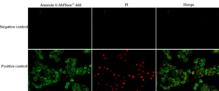
Fig.1 Annexin V-AbFluor™ 488 Apoptosis Detection Kit was used to detect the apoptosis effect of HeLa cells after apoptosis induction
After Annexin V-Abfluor ™ 488 labeling, the necrotic cells and apoptotic cells in early stage and late stage were accurately identified.
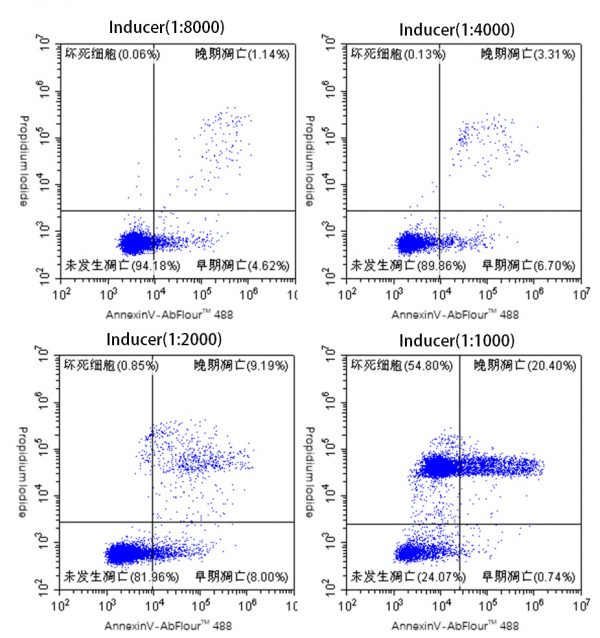
Fig.2 Flow cytometry was used to detect the apoptosis rate by different concentrations of inducer A and B.
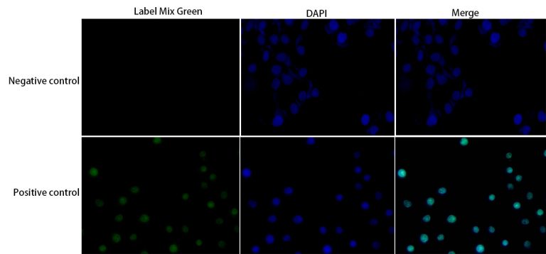
In the detection of apoptosis by TUNEL assay, deoxyribonucleotide terminal transferase (TdT) was used to label the deoxyribonucleotide – luciferase conjugates to the 3′- terminus of the DNA gap, and apoptosis was visualized.
Fig.3 TUNEL Apoptosis Detection Kit (Green Fluorescence) was used to detect the apoptosis effect of HeLa cells after apoptosis induction.
| CAT# | Product name | classification |
| KTB1600 | CheKine™ Reduced Glutathione (GSH) Colorimetric Assay Kit | Cell metabolism |
| KTB1610 | CheKine™ Glutathione Oxidized (GSSG) Colorimetric Assay Kit | Cell metabolism |
| KTB1620 | CheKine™ Glutathione Reductases (GR) Colorimetric Assay Kit | Cell metabolism |
| KTB1630 | CheKine™ Glutathione S-Transferase (GST) Colorimetric Assay Kit | Cell metabolism |
| KTB1640 | CheKine™ Glutathione Peroxidase (GSH-Px) Colorimetric Assay Kit | Cell metabolism |
| KTB1650 | CheKine™ Thioredoxin Reductase (TrxR) Colorimetric Assay Kit | Cell metabolism |
| KTB1660 | CheKine™ Thioredoxin Peroxidase (TPX) Colorimetric Assay Kit | Cell metabolism |
Fig.1. WB experiment of β-Actin expression, the samples are Rat brain (lane 1), HeLa cell lysate (lane 2), Mouse brain (lane 3) each 10 ug, the primary antibody is β-Actin mouse monoclonal antibody ( 1C7) (A01010, 1:5000), the secondary antibody is goat anti-mouse IgG (A21010, 1: 10000).
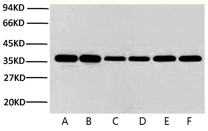
Fig.2. WB experiment of GAPDH, the samples are Hela (lane 1), rat brain (lane 2), Mouse brain (lane 5) each 10 ug, the primary antibody is GAPDH mouse monoclonal antibody (2B5) (A01020, 1 : 10000), the secondary antibody is goat anti-mouse IgG (A21010, 1: 10000).
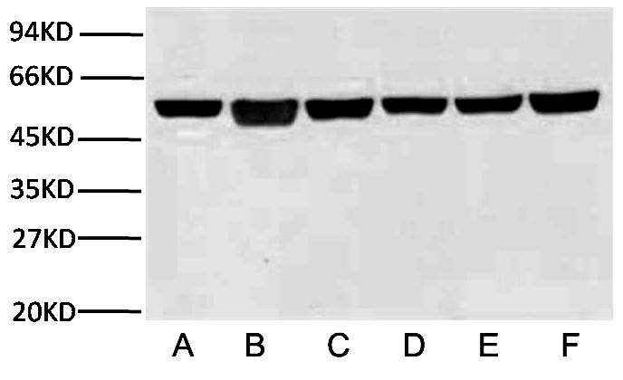
Fig.3. WB experiment of β-Tubulin, the samples are Rat brain (lane 1), A549 cell (lane 2), Mouse brain (lane 3) each 10 ug, the primary antibody is β-Tubulin mouse monoclonal antibody (2B5) (A01030, 1:10000), the secondary antibody is goat anti-mouse IgG (A21010, 1:10000).
 活細胞和死細胞雙染色套組 | CCK-8細胞增殖和細胞毒性試劑盒 | LDH細胞毒性測定試劑盒 | 細胞衰老檢測試劑盒(β-半乳糖苷酶 | 細胞增殖EdU Image試劑盒 | Abbkine 細胞膜染色 | 活細胞Lysosome染色