【SERVICEBIO 組織染色劑】
番紅O (Safranin O)染色試劑盒- ( 軟骨組織 ) | Safranin Solid Green Dye Servicebio貨號G1053-100ML 100 mL×2
番紅O (Safranin O)染色試劑盒- ( 軟骨組織 )
Safranin Solid Green Dye for Soft Bone Tissue Safranin O-Fast Green Staining 番紅固體綠色染料/番紅 O-快速綠色染色(軟骨組織)
產品介紹:
番紅O (Safranin O)是一種能與多種陰離子(multiple anions)結合的陽離子染料(cationic dye)。它能與硫酸軟骨素(chondroitin sulfate )、硫酸角蛋白( keratin sulfate)等軟骨組織中的多醣陰離子基團(polysaccharide anion groups)結合,使其呈紅色。而且番紅O (Safranin O)的顏色大約與陰離子的濃度成正比,可以間接反映軟骨基質中蛋白多醣的含量和分佈。如果軟骨受損,軟骨中的糖蛋白就會丟失,呈現出番紅O (Safranin O)的淺染色或無染色。酸性染料固綠與組織的嗜酸性成分結合,使組織呈現綠色或藍色。軟骨(Cartilage )和骨通過番紅-堅綠染色來區分。本品骨組織番紅固綠染料溶液(bone tissue saffron fast green dye solution),番紅染料溶液( saffron dye solution )濃度為0.2%,固綠染料溶液濃度為2%。適用於骨組織染色。染色後軟骨呈紅色至鮮紅色,成骨(osteogenesis)呈綠色至藍綠色。

| Component Number | Component | G1053-100ML |
| G1053-1 | Safranin O solution | 100 mL |
| G1053-2 | Fast-Green solution | 100 mL |
Assay Protocol / Procedures
1. Paraffin sections were dewaxed to water: sections were successively dewaxed by xylene for 10 min, followed by fresh xylene for 10 min, absolute ethanol for 5min, fresh absolute ethanol for 5min, 90% ethanol for 5min, 75% ethanol for 5min, and washed with tap water.
2. Fast green staining: The sections were stained with fast green staining solution for 1-5 min. The excess staining solution was washed with tap water until the cartilage was colorless. The sections were rapidly treated with 1% hydrochloric acid for 10-15 s, and then washed with tap water.
3. Saffron staining: Sections were stained with saffron staining solution of bone tissue for 5-10 s, and then rapidly dehydrated by four cylinders of absolute ethanol for 3-5 s each time. After the fourth dehydration, microscopic examination showed that the cartilage was red and the background was colorless.
4. Transparent sealing: Sections were transparent through xylene for 5 min, then transparent through fresh xylene for 5 min, and then sealed with neutral gum.
5. Microscopic examination, image acquisition and analysis.
Note: Prepare your own xylene, gradient ethanol, neutral gum, etc.
Note
1. The section should not be stained with saffron for too long, otherwise it is difficult to differentiate, resulting in mixed color with fast green. If you need to re-stain, you can soak in tap water to remove the fast green color, acidic differentiation solution (recommended G1039) to remove the saffrane color, and then stain again after all the colors are decolorized.
2. The excess dye can be removed by washing after fast green staining. Under the microscope, the bone formation is obviously green, and the cartilage is very light or colorless.
3. Wear a lab coat and disposable gloves during operation.
油紅O溶液 | Oil Red O Solution中性脂質染色劑 Servicebio貨號G1015-100ML

產品介紹:
油紅O又稱蘇丹紅5B(Sudan red 5B),是一種脂溶性偶氮染料(fat soluble azo dye)。這種染料可以特異性地染色細胞(cells)或組織(tissues)中的甘油三酯等中性脂質(neutral lipids),但對磷脂(phospholipids)和類固醇(steroids)的染色較弱。基本原理是油紅O(oil red O)溶於脂類(lipids),使脂類(lipids)呈紅色至橙紅色(orange red)。
本品飽和油紅O染料溶液(saturated oil red O dye solution)為油紅O飽和溶液(oil red O),臨用前用蒸餾水稀釋後用於組織切片(tissue sections)或細胞染色。染色後,組織中的脂肪滴(fat drops)呈紅色(red)至橙紅色。 可搭配脂肪專用固定溶劑使用 脂肪專用固定液試劑盒 | Special Fixative Solution for Fat Servicebio貨號G1119-250ML
Oil red O, also known as Sudan red 5B, is a fat soluble azo dye. This dye can specifically stain neutral lipids such as triglycerides in cells or tissues, but it is weak for phospholipids and steroids. The basic principle is that oil red O dissolves in lipids to make lipids red to orange red.
The saturated oil red O dye solution of this product is the saturated solution of oil red O. It can be used to dye tissue sections or cells after being diluted with distilled water before use. After dyeing, the fat drops in the tissues are red to orange red.
Assay Protocol
l Preparation of working solution: before use, (6:4) six portions of saturated oil red O dye solution and four portions of distilled water shall be fully mixed, placed in a 60-70 ℃ water bath for 30 minutes, and then filtered with qualitative filter paper after natural cooling to obtain oil red O working solution. It needs to be prepared and used now. In addition, 60% isopropanol is required.
l Sample preparation
- For cells: aspirate the cell culture medium, slowly add PBS (G4202 is recommended) to the edge of the orifice plate to simply clean the cells. Add 4% paraformaldehyde fixative (G1101 is recommended) and fix it at room temperature for 8-10 min, and rinse it twice with PBS.
- For frozen sections: take out the sections from – 20 ℃ and let them stand at room temperature for 5-10 min to recover to normal temperature.
l Dyeing steps
- For cells
(1) Add a small amount of 60% isopropyl alcohol into the pore plate to cover the cells for 15-20 seconds, and then suck out 60% isopropyl alcohol and dry the water slightly.
(2) Add oil red O working solution to the orifice plate to cover the cells, and dye them at room temperature in dark for 30 min to remove the dye.
(3) Add 60% isopropanol for rapid differentiation for 3-5 seconds, wash with pure water for 3 times, and each time for 5 minutes.
(4) (Optional) Add hematoxylin dye solution (G1004 is recommended) to dye the nucleus, wash with water, turn blue and then wash with water.
(5) PBS was added to cover the cells and observed under microscope. In case of cell climbing, glycerol gelatin film sealant (G1402 is recommended) can be used for slide mounting.
- For frozen sections
(1) Frozen sections recovered to room temperature were gently immersed in oil Red O working solution and stained for 8-10 min (covered to avoid light).
(2) The sections were taken out, stayed for 3 s, and then immersed in two cylinders of 60% isopropanol for differentiation for 3-5 s.
(3) Sections were immersed in two cylinders of pure water for 10 s each time.
(4) (Optional) The sections were immersed in hematoxylin dye solution to stain the nuclei, washed with water, then returned to blue and washed again. After slightly drying, glycerin gelatin was added to mount the slide.
Note:
- If it is a frozen section of fresh tissue, the section should be fixed before staining.
- Oil Red O working fluid must be prepared and used at present. The heating process must be sealed to prevent solvent volatilization.
- During the whole operation, pay attention to the gentle action to avoid fat loss or displacement.
- Samples stained with oil Red O cannot be stored for a long time, and should be observed and photographed as soon as possible.
- When using glycerin gelatin to mountslides, attention should be paid to avoid bubbles as far as possible. If bubbles are not allowed to press the glass slide or forcibly tear the cover glass after mounting the slides, it will cause fat displacement. The slide can be immersed in warm water at 50-60 °C to allow the cover slide to fall off and then re-mount the slide.
- For your safety and health, please wear a lab coat and disposable gloves during operation.
Wright-Giemsa 染色劑 | 血液(blood)和骨髓( bone marrow )塗片的染色劑貨號G1007-250ML 250ml+500ml
產品介紹:
賴特氏染色劑(Wright-Giemsa Staining Kit)是促進血細胞類型分化的血液染色劑。它通常是曙紅和亞甲藍染料的混合物。它主要用於對外周血塗片,尿液樣本和骨髓抽吸物進行染色,並在光學顯微鏡下進行檢查。在細胞遺傳學中,它用於染色染色體以促進綜合症和疾病的診斷。
Wright-Giemsa Stain 主要用於血液(blood)和骨髓( bone marrow )塗片的染色。 它是根據羅曼諾夫斯基染色技術(Romanowsky Stain technology)的原理進行修飾的。 細胞的染色過程是染料滲入被染物並保留在其內部的過程,既有物理吸附作用,也有化學親和作用。 由於化學性質不同,各種細胞和細胞成分對萊特-姬姆薩染色液中的酸性染料(伊紅/eosin)和鹼性染料(亞甲藍/methylene blue)的親和力不同。
本品包括兩種溶液,A液含0.2% Wright和0.1% Giemsa,B液為磷酸鹽緩衝液。 標本塗片經本品染色後,各種細胞呈不同顏色,其中紅血球細胞呈淡紅色,白細胞血漿顆粒清晰,呈現出各種細胞特有的顏色。 嗜酸性粒細胞胞質顆粒呈紫紅色,嗜鹼性粒細胞胞質顆粒呈藍色,單核細胞和淋巴細胞呈藍色。 因此,本產品可達到區分各種細胞形態特徵的目的。
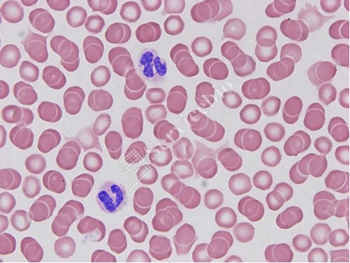
Wright – Giemsa Stain is mainly used for staining blood and bone marrow smears. It is modified based on the principle of Romanowsky Stain technology. The dyeing process of cells is a process that the dye penetrates into the dyed material and remains inside it,w hich involves both physical adsorption and chemical affinity. Due to their different chemical properties, various cells and cell components have different affinities for acid dyes (eosin) and basic dyes (methylene blue) in Wright-Giemsa staining solution.
This product includes two kinds of solution , solution A contains 0.2% Wright and 0.1% Giemsa, and solution B is a phosphate buffer solution. After the specimen smear is stained with this product, various types of cells show different colors, among which the red blood cells are light red, the white blood cell plasma particles are clear, and show the unique colors of various cells. The cytoplasmic granules of eosinophils are purplish red, the cytoplasmic granules of basophils are blue, the monocytes and lymphocytes are blue. Therefore, this product can achieve the purpose of distinguishing various cell morphological characteristics.
| Component Number | Component | G1007-20ML | G1007-2 5 0ML |
| G1007-1 | Wright-Giemsa S olution A | 20 m L | 250 mL |
| G1007- 2 | Wright – Giemsa S olution B | 40 m L | 2×250 mL |
| Instruction | 1 | ||
革蘭氏染色試劑Gram Staining Kits | Servicebio貨號G1065-20ML (4x20ml)
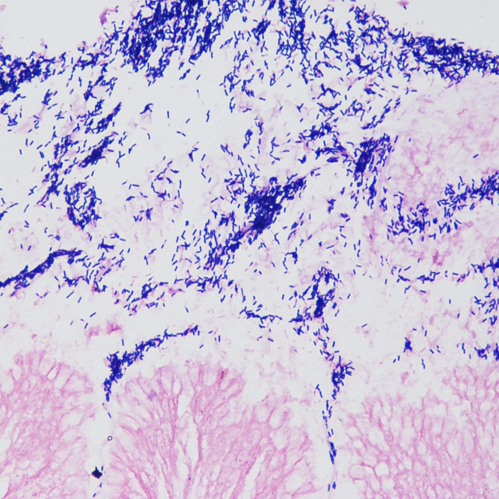
革蘭氏染色(Gram staining)是微生物學(microbiology)中廣泛應用的一種鑑別染色方法,可用於區分革蘭氏陽性菌(Gram-positive bacteria , G+)和革蘭氏陰性菌(G-)
產品介紹:
革蘭氏染色(Gram staining)基本原理是基於細菌細胞壁(bacterial cell walls)的不同化學成分(chemical components)。 在用結晶紫(crystal violet)初步染色並用碘媒(iodine)染後,細菌在細胞壁中形成不溶於水的結晶紫-碘複合物(water-insoluble crystal violet-iodine complexes)。革蘭氏陽性菌(Gram-positive bacteria)細胞壁(cell walls)厚,不含脂質(lipids),富含肽聚醣(peptidoglycan),交聯形成緻密的網格結構(dense grid structure)。當用乙醇(ethanol)處理時,肽聚醣網收縮,可以阻擋結晶紫-碘絡合物。它保留在細胞壁中,使其呈現結晶紫的紫色。另一方面,革蘭氏陰性細菌具有薄的細胞壁、低肽聚醣含量和鬆散的交聯。當接觸乙醇時,富含脂質的外膜(lipid-rich outer membrane)溶解,細胞壁(cell wall)出現大的空隙,結晶紫-碘絡合物(crystal violet-iodine complex)流出,乙醇脫色(decolorization)後細胞變為無色。此時用番紅(safranin)複染(counterstaining),革蘭氏陰性菌(Gram-negative bacteria)會被染成紅色。

革蘭氏染色液含有四種成分,即草酸銨結晶紫染色液(ammonium oxalate crystal violet staining solution)20ml、1%碘液(iodine solution)20ml、脫色液(destaining solution)20ml和品紅染色液(fuchsin staining solution)20ml。染色後,革蘭氏陽性菌(gram-positive bacteria)呈紫色至藍紫色(blue-purple),革蘭氏陰性菌(gram-negative bacteria)呈紅色(red)。
Assay Protocol / Procedures
一、石蠟切片脫蠟(Dewax)去水;將細菌塗片(bacterial smear)自然晾乾或用酒精燈加溫晾乾固定。. Dewax the paraffin section to water; dry the bacterial smear naturally or warm it with an alcohol lamp to dry and fix it.
二、初染:滴加結晶紫染色液(crystal violet staining solution)覆蓋樣品10-30s,水洗,甩乾。Initial dyeing: Add crystal violet staining solution dropwise to cover the sample for 10-30 s, wash with water, and spin dry.
三、媒染:滴加1%碘溶液(iodine solution)覆蓋樣品1-1.5分鐘,水洗,甩乾。Mordant dyeing: Add 1% iodine solution dropwise to cover the sample for 1-1.5 min, wash with water, and spin dry.
四、脫色(Decolorization):滴加脫色液(decolorization solution)沖洗玻片一端脫色的組織,直至流下的脫色液不再呈紫色,立即用水洗去脫色液。注意控制脫色時間。Decolorization: dropwise add decolorization solution to rinse the tissue from one end of the slide for decolorization, until the decolorization solution that flows down no longer appears purple, and immediately wash with water to remove the decolorization solution. Take care to control the destaining time.
五、複染(Counterstaining):滴加品紅染色液(Fuchsin staining solution )20-60 s,然後水洗。用濾紙吸乾邊緣。Counterstaining: add Fuchsin staining solution dropwise for 20-60 s, then wash with water. Blot the edges with filter paper.
六、細菌塗片(Bacterial smears)可直接用顯微鏡觀察。 60 ℃烘乾後,組織切片在無水乙醇(absolute ethanol)中快速脫水 3 次,時間分別為 1 s、3 s 和 5 s。然後,用二甲苯(xylene)透明化5分鐘,最後用中性樹膠(neutral gum)密封以進行顯微鏡檢查。Bacterial smears can be observed directly by microscopy. After drying at 60 °C, the tissue sections were rapidly dehydrated in absolute ethanol for three times, 1 s, 3 s, and 5 s, respectively. Then, it was transparentized with xylene for 5 min, and finally sealed with neutral gum for microscopic examination.
| Component number | Component | G1401-20ML |
| G1065-1 | Ammonium oxalate crystal violet staining solution | 20 mL |
| G1065-2 | 1% iodine solution | 20 mL |
| G1065-3 | destaining solution | 20 mL |
| G1065-4 | Fuchsin staining solution | 20 mL |
| Instruction | 1 | 1 |
Masson’s three-color staining | 結締組織染色Servicebio貨號G1006-100mL(6×100 mL)
產品介紹:
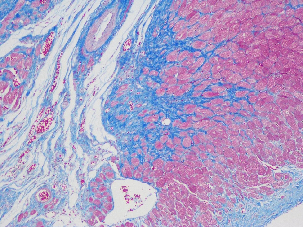
Masson’s三色染色法(Masson’s Trichrome Stain),是最常被使用的染色法之一,又稱Masson’s染色 (Masson 膠原纖維三色染色液),是比較經典的膠原纖維染色法(collagen fiber staining method)。該方法利用結締組織(connective tissues)中陰離子生物染料(anionic biological dyes)和分子量(molecular weights)的不同滲透性。分子量小的染料很容易穿透結構緻密、滲透性低的組織,而分子量大的染料只能進入結構疏鬆、滲透性好的組織,因此不同的組織成分呈現出不同的顏色。傳統的膠原纖維 Collagen fiber染色技術,主要用於膠原纖維與肌肉纖維的鑑別染色,尤其經常被用以觀察組織的纖維化(fibrosis)。故常被用於腎臟、肺臟與肝臟等組織的纖維化判定依據與投藥改善的評估方式之一。

本品為試劑盒,主要成分如下:A液為2.5%重鉻酸鉀(2.5% Potassium dichromate),B液、C液等體積混合為魏格鐵蘇木精染液(Weigert iron hematoxylin dye solution),D液為麗春紅酸品紅(Ponceau acid fuchsin),E液為1 %磷鉬酸溶液(phosphomolybdic acid solution),F液為2.5%苯胺藍溶液(2.5% aniline blue solution)。結締組織切片(connective tissue sections stain)經Masson’s三色染色液染色後,膠原纖維(collagen fibers)呈天藍色至亮深藍色,肌纖維(muscle fibers)、細胞質(cytoplasm)、纖維素(cellulose)、角質白(keratinous white)呈紅色至紫紅色,紅血球細胞(red blood cells)呈淡紅色。
| Component Number | Component | G1006-20ML | G1006-100ML |
| G1006-1 | Masson A solution | 30 mL | 100 mL |
| G1006-2 | Masson B solution | 20 mL | 100 mL |
| G1006-3 | Masson C solution | 20 mL | 100 mL |
| G1006-4 | Masson D solution | 20 mL | 100 mL |
| G1006-5 | Masson E solution | 20 mL | 100 mL |
| G1006-6 | Masson F solution | 20 mL | 100 mL |
Assay Protocol
- Section preparation: paraffin sections are deparaffinized to water; frozen sections stored at -20°C need to be allowed to stand and return to room temperature.
- Soak the slices in Masson Asolution at room temperature overnight (about 15 hours).
- Soak the slices in Masson A solution and incubate in a 65° oven for 30 mins. Wash with tap water for 30 s until the yellow color on the tissue fades. At the same time, put Masson D liquid and Masson F liquid in a 65° oven to preheat.
- Mix Masson B solution and Masson C solution in equal volume ( prepared for current use, not pre-prepared and preserved ), slice into the mixed solution and soak for 1 min, then rinse with running water.
- Put the slices into 1% hydrochloric acid alcohol (concentrated hydrochloric acid: absolute ethanol = 1:100) for differentiation for about 1 min,until the nucleus was gray-black, and the background was almost colorless or light gray.
- Rinse with tap water and drain the excess water on the slices. Dip the slices into Masson D solution for 6 minutes. At this time, the tissues will appear bright red. If the red is too light, the staining time can be prolonged.
- Drain the slices slightly (not dry slices), and soak in Masson E solution for about 1 min. This step is for differentiation. It is enough to differentiate until the collagen fibers are light red and the fibers are red. The time of E solution can be adjusted according to the degree of staining, usually 1-2 min.
- After the section is slightly drained of Masson E solution, without water washing, it is directly stained into Masson F solution for 2-30 s.
- The slices are rinsed and differentiated in three consecutive tanks with 1% glacial acetic acid, each tank is about 8 seconds.
- The slices are dehydrated in successive three cylinders of absolute ethanol for about 5 s, 10 s, and 30 s. Then two cylinders of n-butanol were dehydrated for 30 s and 2 minutes in sequence, and finally two cylinders of xylene were used to be transparent, 5 minutes each time, and the sheets were sealed with neutral gum.
蘇木精-伊紅染色試劑 (H&E Staining Kit) | H&E Staining Kit Servicebio貨號G1005-500ML (2X500ML)
H&E蘇木精-伊紅染色
HE stain 又稱蘇木素-伊紅染色,或「H&E染色」H&E 染色試劑盒 (H&E Staining Kit, Hematoxylin and Eosin) 旨在用於組織學(histology)和細胞學(cytology)應用。H&E染色組織及細胞結構清晰,為最廣泛應用於組織切片形態觀察的染色方法之一,這種染色方法的基礎是組織結構對不同染料的結合程度不同。蘇木素是鹼性染料,可以將嗜鹼性結構染成藍紫色。被蘇木素著色的結構本身為酸性,具有嗜鹼性(Basophilic),鹼性結構通常包括含有核酸的部分,如核糖體、細胞核及細胞質中富含核糖核酸(RNA)的區域等。伊紅是酸性染料,粉紅色。可以將大多數細胞的細胞質染成紅色。被伊紅著色的結構本身為鹼性,具有嗜酸性(Acidophilic)。不易被蘇木素和伊紅著色的結構具有嗜中性(Neutrophilic)。H&E 染色試劑盒包括Hematoxylin Stain solution, 500ml及Eosin stain solution, 500ml。

H&E Staining Kit (Hematoxylin and Eosin) is intended for use in histology and cytology applications. Included in this kit is a newly formulated Eosin that provides the benefits of a traditional alcoholic formula with significant improvements in usability. Advantages include lower evaporation rate, better color patterns, reduced tendency to spill over container, hands, and countertops, and improved surface tension to remain on tissue section. The Hematoxylin produces crisp, intense blue nuclei providing optimal contrast to the Eosin Y stained cytoplasm. After tissue sections or cells are stained, the nuclei are blue, the cytoplasm are light pink, collagen are pink, muscle are pink to rose, and erythrocytes are pink to red.

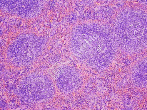
Mouse Ovarium Histology and Morphology
產品介紹:
H&E 染色試劑盒 (H&E Staining Kit, Hematoxylin and Eosin) 。該套件中包含一種新配製的曙紅(Eosin),它具有傳統酒精配方的優點,並顯著提高了可用性。優點包括較低的蒸發率(evaporation rate)、更好的顏色圖案(better color patterns)、減少溢出容器(spill over container)、手和工作檯面的可能性,以及提高表面張力以保留在組織切片上。蘇木精(Hematoxylin)產生清晰、強烈的藍色細胞核(nuclei),與伊紅 Y 染色(Eosin Y stained)的細胞質(cytoplasm)形成最佳對比。組織切片(tissue sections)或細胞(cells)染色後,細胞核(nuclei)呈藍色,細胞質(cytoplasm)呈淡粉色,膠原蛋白(collagen)呈粉紅色,肌肉(muscle)呈粉紅色至玫瑰色,紅血球細胞(erythrocytes)呈粉紅色至紅色。
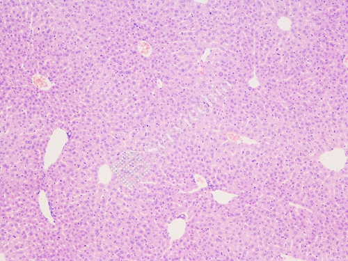
Mouse(Liver) Histology and Morphology
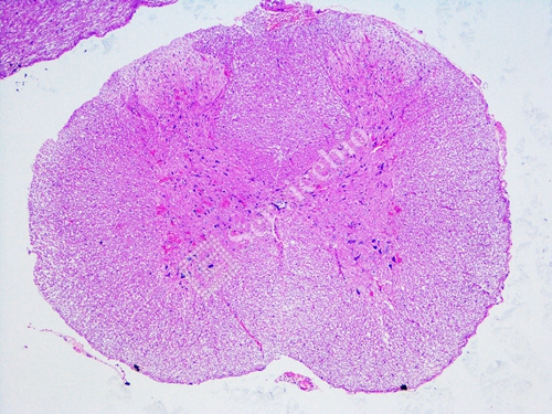
Rat (Spinal Cord) Histology and Morphology
Directions for Use
- Deparaffinize sections if necessary and hydrate to distilled water.
- Rinse slide in distilled water.
- Stain slide in Hematoxylin, Mayer’s for 3-5 minutes.
- Rinse slide in running tap water.
- Apply Differentiation Reagent for 2-5 seconds, rinse slide in running water.
- Apply Bluing Reagent for 30 seconds.
- Rinse in distilled water.
- Dip slide in 85% alcohol for 3-5 min, and then 95% alcohol for 3-5 min, and blot excess off.
- Apply adequate Eosin Y Solution (Alcoholic) to completely cover tissue section to excess and incubate for 2-5 minutes.
- Rinse slide using absolute alcohol.
- Dehydrate slide in 3 changes of absolute alcohol.
- Clear slide and mount in synthetic resin.
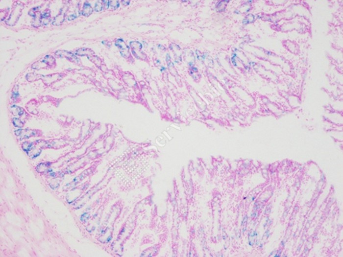
Mouse (Cerebellum) Histology and Morphology
核固紅溶液 | Nuclear Fast Red Stain核複染溶液 Servicebio貨號G1035-100ML
產品介紹:核固紅(Nuclear fast red , CAS#6409-77-42)。是一種優化的核複染溶液,可產生強烈的核染色效果。核固紅(Nuclear fast red)它是一種陰離子染料,可以通過離子鍵(ionic)或氫鍵(hydrogen)與帶負電的原子核( negatively charged nucleus)結合,使原子核染色。與蘇木精相比,核固紅是一種快速染色程序,只需一步即可在五分鐘內對細胞核進行染色。核固紅染色劑(nuclear fast red stain)的有效成分濃度為0.1%,常用於核染色。當組織切片或細胞被染色時,細胞核呈紅色。
Assay Protocol
1. 切片進行脫蠟並水合至蒸餾水 Deparaffinize sections if necessary and hydrate to distilled water.
2. 應用核固紅溶液(增強穩定性)並孵育 5-10 分鐘Apply Nuclear Fast Red Solution (Enhanced Stability) and incubate for 5-10 minutes.
3. 沖洗Rinse with running water.
4. 分級酒精(70%-80%-95%-100%)脫水Dehydrate through graded alcohols (70%-80%-95%-100%).
5. Clear, and mount in synthetic resin.