衰老細胞β-半乳糖苷酶染色試劑盒 | Aging Cell β-galactosidase Staining Kit Servicebio貨號G1073-100T
| 代理廠牌: | |
| 原廠連結: | |
| 相關下載: |
衰老細胞β-半乳糖苷酶染色試劑盒 | Aging Cell β-galactosidase Staining Kit Servicebio貨號G1073-100T (1ml為一個反應單位)

產品介紹:
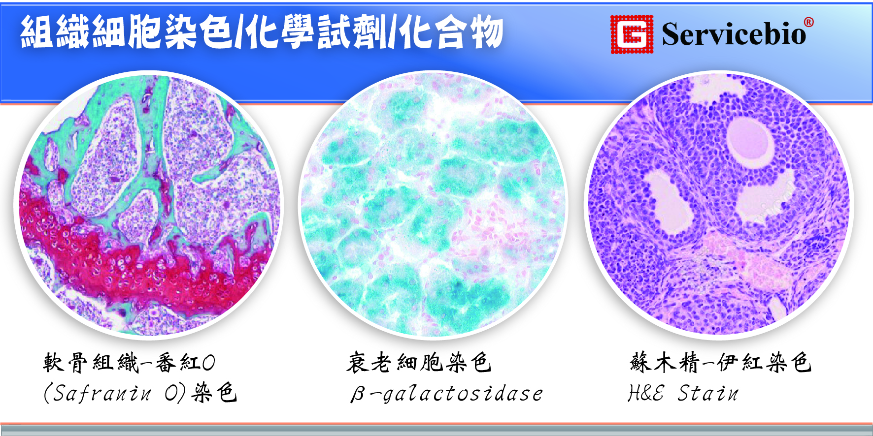
大多數正常細胞的分裂能力(division ability)是有限的。當它們不能分裂(divide)時,就會進入衰老狀態(senescence),稱為細胞衰老(cell senescence)。細胞衰老(Cell senescence)是細胞控制其生長潛能(growth potential)的保證機制,一般指複製性衰老(replicative senescence)。正常細胞在分裂次數有限後停止分裂,並發生不可逆的生長停滯(irreversible growth arrest)。此時,細胞還活著,但細胞形態(cell morphology)和生理代謝活動(physiological metabolic activity)發生了顯著變化,通常表現為細胞體積變大和與衰老相關的β-半乳糖苷酶(β-galactosidase)激活。 β-半乳糖苷酶(β-galactosidase)是細胞溶酶體(cell lysosomes)中的一種水解酶(hydrolytic enzyme)。它通常在 pH 4.0 時有活性,但在衰老細胞(senescent cell)中它在 pH 6.0 時有活性。本試劑盒正是基於這一現象和原理對衰老組織或細胞進行染色,以對抗與衰老相關的β-半乳糖苷酶(β-galactosidase)活性水平的上調。具體反應原理是以X-Gal為底物,衰老細胞(senescent cell)特異性β-半乳糖苷酶催化底物生成藍色產物,在細胞質中表現為藍色沉澱物,在光照下可觀察到顯微鏡。按每個樣品染色液用量1 mL計算,該試劑盒可完成100個樣品的染色。

Fig.1 WI-38 cells were stained with β-galactosidase kit. The left picture shows senescence WI-38 cells without division and proliferation ability but still alive. After staining, the positive staining cells were more than 95%. The image on the right shows newly resuscitated WI-38 cells (early passage) with less than 3 passages, and no obvious positive cells after staining.
| Component Number | Component | G1073 |
| G1073-1 | β-galactosidase staining fixation solution | 100 mL |
| G1073-2 | β-galactosidase staining solution A | 100 mL |
| G1073-3 | β-galactosidase stain B | 1.2 mL |
| G1073-4 | DMF | 5 mL |
| G1073-5 | X-Gal(powder) | 100 mg |
Assay Protocol
l Preparation of reagents
- Prepare your own PBS buffer (G4202 recommended).
- 100 mg X-Gal powder was fully dissolved and mixed with 5 mL DMF (dimethylformamide), and then divided into 1.5 mL clean centrifuge tubes, 0.5 mL for each tube, and stored at -20℃ away from light. Avoid repeated freezing and thawing.
- Preparation of β-galactosidase staining solution according to the proportion in the table below. For cells cultured in 6-well plates, 1.0-1.5 mL of staining working solution is required per well, and for 12-well plates, 0.5-1.0 mL of staining working solution is required per well. The staining solution was prepared according to the sample size to avoid waste.
| Component | Volume |
| β-galactosidase staining solution A | 940 μL |
| β-galactosidase stain B | 10 μL |
| X – Gal solution | 50 μL |
| Total Volume | 1 mL |
![]()
Staining procedure
貼壁細胞 For adherent cells
(1) The cultured cells (or cell crawling sheets) in 6-well plates were aspirated and the cell culture medium was removed, washed twice with PBS, and 1 mL β-galactosidase staining fixing solution was added, and the cells were fixed for 15 min at room temperature.
吸去6孔板中培養的細胞(或細胞爬片)並吸去細胞培養基,用PBS洗滌2次,加入1 mL β-半乳糖苷酶染色固定液,室溫固定細胞15分鐘。
(2) The fixed solution was discarded, and the cells were washed with PBS for 3 times, 2 min each time.
棄去固定液,用PBS洗滌細胞3次,每次2分鐘。
(3) PBS was removed by suction with a pipette, and 1 mL of β-galactosidase staining working solution was added to each well and incubated at 37℃ for 2 h to overnight. Note: Do not incubate in carbon dioxide incubator at 37 ° C. During the staining period, the color development should be observed in time. If the expression of β-galactosidase in the sample is high, the staining can be completed within a few hours. If β-galactosidase expression was low, the incubation time should be extended appropriately, during which the 6-well plate should be sealed with plastic wrap or parafilm to prevent liquid evaporation from affecting the staining results.
用移液管吸去PBS,每孔加入1 mL β-半乳糖苷酶染色工作液,37℃孵育2小時至過夜。注意:染色過程中請勿置於37℃二氧化碳培養箱中培養。染色期間應及時觀察顯色情形。如果樣品中β-半乳糖苷酶表現量較高,染色可在數小時內完成。若β-半乳糖苷酶表現量較低,則應適當延長孵育時間,並以保鮮膜或石蠟膜封好6孔板,防止液體蒸發影響染色結果。
(4) Under the ordinary light microscope, the staining solution was removed after the positive cells developed color. If nuclei need to be counterstained, add a small amount of nucleosinophilic red dye solution (G1035 is recommended) to the well plate to cover the cells and stain at room temperature for 3 min, remove the staining solution, and wash with PBS several times.
在普通光學顯微鏡下,待陽性細胞顯色後吸去染色液。如需細胞核複染,則在孔板中加入少量核苷紅染料溶液(建議使用G1035)覆蓋細胞,室溫染色3分鐘,吸去染色液,用PBS洗滌數次。
(5) 2 mL PBS was added to cover the cells and the staining was completed. The sample could be stored at 4℃ for 1 week. Or add 70% glycerol to cover the cells, 4℃ can be stored for a long time. If it is the cell climbing sheet, the climbing sheet can be fully dried, xylene transparent after dropping neutral gum seal sheet, can be stored for a long time.
冰凍切片(For frozen sections)
(1) Rewarm frozen sections at room temperature for 10 min. Circle the tissue with tissue strokes.
將冰凍切片室溫復溫10分鐘。用組織筆畫圈出組織。
(2) A proper amount of β-galactosidase staining fixing solution was added to the tissue to completely cover the tissue, and the solution was fixed at room temperature for 20 min.
將適量β-半乳糖苷酶染色固定液加入組織上,使其完全覆蓋組織,室溫固定20分鐘。
(3) The tissue sections were soaked and washed in PBS for 3 times, 5 min each time.
將組織切片用PBS浸泡清洗3次,每次5分鐘。
(4) The sections were placed in a wet box to avoid light, and an appropriate amount of β-galactosidase staining solution was added to the tissue to completely cover the tissue. The wet box was incubated at 37℃ and the color development was observed under a microscope every 2 h. If no color development was observed, the culture was continued until the senescent cells on the tissue showed color. If the sample is to be incubated overnight, a sufficient amount of β-galactosidase staining solution should be added to prevent the staining solution from evaporating and drying the tablets.
將切片置於避光濕盒中,將適量β-半乳糖苷酶染色液加入組織上,使其完全覆蓋組織。將濕盒置於37℃培養箱中,每2小時在顯微鏡下觀察顯色情形。若未觀察到顯色,則繼續培養至組織上老化細胞顯色。若需過夜培養,則應加入足量的β-半乳糖苷酶染色液,以防止染色液蒸
(5) After the tissue developed color, the staining solution was removed, and the sections were immersed in PBS and washed twice, and then immersed in pure water and washed twice.
待組織顯色後,吸去染色液,將切片浸入PBS中洗滌2次,再浸入純
(6) (optional) Add nuclear solid red dye solution (G1035 is recommended) for 3 min and wash for 3 times.
最佳化加入核固紅染液(建議使用G1035)3分鐘,洗滌3次。
(7) The slices were dehydrated with absolute ethanol for 2 times, then transparent with xylene for 5 min each time, and then sealed with neutral gum drop.
切片用無水乙醇脫水2次,再用二甲苯透明,每次5分鐘,最後用中性膠封片。
- Staining results
The cytoplasm of senescent cells is scattered blue.
Note:
- X-Gal solution should be thawed and mixed completely at room temperature before use.
- β-galactosidase staining solution A and B should be restored to room temperature in advance before use, and the prepared staining solution should be thoroughly mixed without precipitation before use.
- The β-galactosidase staining reaction of senescent cells is dependent on specific pH conditions, so it cannot be incubated in a CO2 incubator for color development, otherwise it will affect the pH of the staining solution and lead to staining failure.
- When preparing dyeing solution, please choose consumables made of polypropylene (PP) or glass instead of polystyrene (PS).
- The color development should be observed several times during the 2 h-overnight color development period, too short a time may lead to negative results; too much time can lead to false positives. The chromogenic time is closely related to the amount of β-galactosidase contained in the sample itself.
- Before preparing the staining solution, check the pH value of staining solution A. If it is not 6.0 (which may be changed due to storage conditions), adjust the pH value to 6.0 with HCl or NaOH before use.
- β-galactosidase staining of tissue sections requires high preparation of samples, which should be stored at -80℃ and tested as soon as possible. Because β-galactosidase is very easy to inactivate, improper storage or too long of the sample may lead to enzyme inactivation, then no positive staining.
- Please wear a lab coat and disposable gloves during operation
Servicebio台灣品牌介紹-BRAND/AGENCY/品牌介紹- Servicebio