WST-1細胞存活分析(呈色)套組| WST-1 Cell Proliferation Colorimetric Assay Kit K301-500(abcam ab65473)
| 代理廠牌: | |
| 原廠連結: | |
| 相關下載: |
特點
Cell Proliferation Assay Kits | 貨號K301-500 (abcam ab65473)
Biovision 系列細胞存活 | 細胞毒性分析套組 (呈色/螢光/冷光)
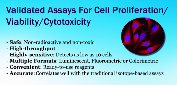
WST-1 Cell Proliferation Colorimetric Assay Kit WST1細胞活性分析呈色套組 (貨號K301-500);快速細胞增殖分析,使用水溶性tetrazolium鹽類的的WST-1作為染劑,利用粒腺體中的脫氫酶還原成橙色產物。WST-1比 起傳統的MTT、XTT或是MTS-base分析要來的更快速且靈敏度更高。| 細胞老化分析套組|Senescence Detection Kit: 細胞老化被認為是抑制癌細胞的機制,因此當細胞老化時雖然細胞會持續存活,但是在這個階段的細胞是不會分裂,也無法以血清及培養基進行細胞繫代培養。Senescence detection kit是偵測切片中組織或細胞上 特定的senescence marker,而這些marker是不會出現在其他的細胞中。|生物冷光細胞毒性分析套組 | Bioluminescence Cytotoxicity Assay Kit: 這套細胞毒殺試劑組是透過受損傷的細胞釋放Adenylate Kinase (AK),利用AK將phosphorylates ADP 轉化為 ATP,產生的 ATP 能夠參與 Luciferase 冷光反應,再透過冷光偵測儀偵測結果,每次分析只要 10 個以上細胞就能分析,而且只要10分就能得到結果。| LDH細胞毒性分析套組 |LDH-Cytotoxicity Assay Kit: 這個試劑組是透過偵測乳酸脫氫酶(lactate dehydrogenase, LDH)活性判斷細胞毒殺現象。LDH存在於所 有細胞中,當細胞破裂時才會釋放至細胞外。使用LDH-cytotoxicity assay kit只需要分光光度計500nm 波長的區段,避閧放射線分析方法,可用於貼附型或懸浮型細胞培養液。操作時間大約30-60分鐘。|死活細胞染色 | Live-Dead Cell Staining Kit: Live-Dead Cell Staining Kit是ready-to-use的試劑,裡面含2種不同波長的染劑,包括染活細胞的Live -dye (綠螢光,Ex/Em = 490/515 nm)以及染死細胞的propidium iodide (PI,紅螢光,Ex/Em = 488/615 nm),細胞經過染色後,能夠直接在螢光顯微鏡下觀察細胞存活的狀況。
產品介紹:
快速細胞增殖分析試劑盒 (The Quick Cell Proliferation Assay Kit , K301) 用於快速,靈敏地定量細胞增殖 ( cell proliferation) 和活力 (viability) 。原理為利用細胞粒腺體脫氫酶 ( mitochondrial dehydrogenases) 將四唑鹽WST-1 ( tetrazolium salt WST-1 ) 裂解成 formazan。活細胞數量增加會導致線粒體脫氫酶 (mitochondrial dehydrogenases) 活性的增加,而形成的formazan dye的量增加。活細胞產生的 formazan dye可以通過多孔分光光度計(微量Reader)通過測量染料溶液在440nm處的吸光度來定量。該測定法可應用於測量生長因子 (growth factors) 於細胞增殖之反應,細胞因子(cytokines) ,促分裂原(mitogens) 和營養素 (nutrients) 等的細胞增殖 ( cell proliferation) 。它還可用於分析細胞毒性化合物,如抗癌藥物和許多其他毒性劑和藥物化合物。新方法非常簡單,無需洗滌,無需收穫,也無需溶解步驟,並且比MTT,XTT或基於MTS的分析更快,更靈敏。整個測定可以在微量滴定板中進行。此產品需由儀器進行定量 ELISA儀器設備請按此。
細胞活性呈色分析套組 | 相關分析
K301 引用文獻
Hsueh, Yu-Sheng et al. (2017) Laminin-Alginate Beads as Preadipocyte Carriers to Enhance Adipogenesis In Vitro and In Vivo, Tissue Eng Part A. 2017 Mar;23(5-6):185-194.
Fang, Y. et al., (2017) IL-33 acts as a foe to MIA PaCa-2 pancreatic cancer, Medical Oncology, Feb.2017, 34(2), 23
Cheng et al., Autophagy modulates endoplasmic reticulum stress-induced cell death in podocytes: A protective role. Experimental Biology and Medicine, Oct 2014; 10.1177/1535370214553772.
Murdoch et al., Glutaredoxin-1 Up-regulation Induces Soluble Vascular Endothelial Growth Factor Receptor 1, Attenuating Post-ischemia Limb Revascularization. J. Biol. Chem., Mar 2014; 289: 8633 – 8644.
Sefton et al., MK-2206, an AKT Inhibitor, Promotes Caspase-Independent Cell Death and Inhibits Leiomyoma Growth.Endocrinology, Nov 2013; 154: 4046 – 4057.