7-AAD (7-aminoactinomycin D) 螢光 DNA 結合染料 | Biotium 貨號40084 (1mg/ml)

| 代理廠牌: | |
| 原廠連結: | |
| 相關下載: |
7-AAD (7-aminoactinomycin D) 螢光 DNA 結合染料 | Biotium 貨號40084 (1mg/ml)
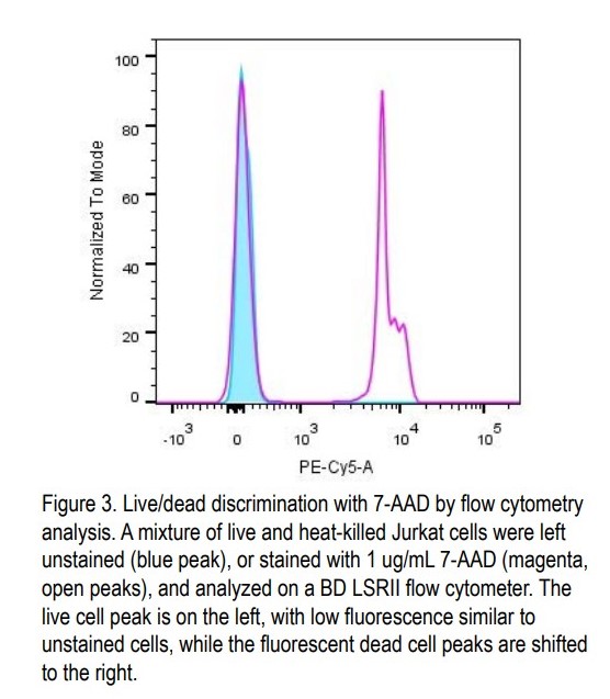
7-AAD(7-氨基放線菌素 D, 7-aminoactinomycin D)是一種膜不可滲透(membrane-impermeant )的螢光 DNA 結合染料,可用於通過流式細胞儀( flow cytometry) 進行活/死細胞區分( live/dead discrimination )和細胞週期分析(cell cycle profiling)。7-AAD 是一種螢光 DNA 結合染料(fluorescent DNA binding dye ),不透膜(membrane impermeant ),因此通常被活細胞( live cells )和早期凋亡細胞( early apoptotic cells)排除,但會染色壞死( necrotic )和晚期凋亡細胞(late apoptotic cells ),膜完整性受損( membrane integrity)。

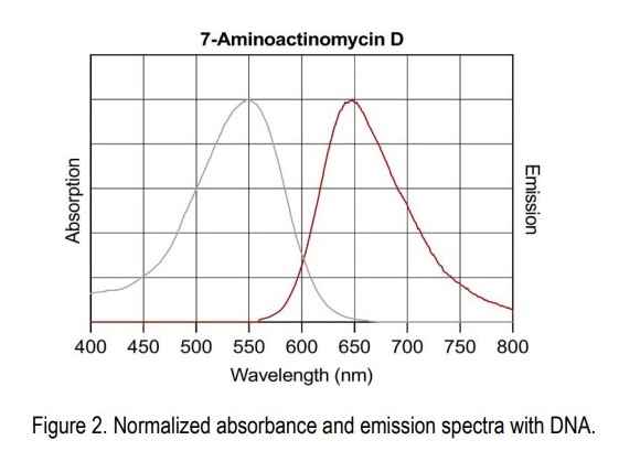
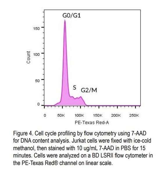
與 DNA 結合的 7-AAD 在激發/發射波長為 546/647 nm 時具有較大的Stokes shift 。染料可以用 488 nm 或 532 nm 激光線激發,並在 PE-Cy®5/PerCP 流式細胞儀通道中檢測。 7-AAD 廣泛用於區分活細胞/死細胞。它還可用於固定和透化細胞,通過使用流式細胞儀進行 DNA 含量分析來進行細胞週期分析。 7-AAD 選擇性地嵌入富含 GC 的 DNA 區域,使其染色可用於染色體顯帶研究。我們提供固體粉末形式的 7-AAD(貨號 40037)或 1 mg/mL DMSO: 水 (1:1) 溶液(貨號40084)。建議使用 1 ug/mL(1:1000 稀釋)的染料來檢測活/死細胞或 10 ug/mL(1:100 稀釋)用於細胞週期分析。
7-AAD (7-aminoactinomycin D) is a membrane-impermeant fluorescent DNA binding dye that is useful for live/dead discrimination and cell cycle profiling by flow cytometry.
- Selective detection of dead cells by flow cytometry in the PE-Cy®5/PerCP channel
- Perform cell cycle profiling by flow cytometry in fixed/permeabilized cells
- λEx/λEm (with DNA) = 546/647 nm (7-AAD can also be excited by the 488 or 532 laser lines)
- Orange red solid soluble in DMF or DMSO
- Store at -20°C and protect from light, especially in solution
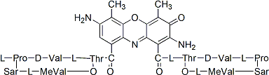
- C62H87N13O16
- MW: 1270.45
建議使用
建議使用 1 ug/mL(1:1000 稀釋)的染料來檢測活/死細胞或 10 ug/mL(1:100 稀釋)用於細胞週期分析
We recommend using the dye at 1 ug/mL (1:1000 dilution) for live/dead discrimination or 10 ug/mL (1:100 dilution) for cell cycle profiling. Example protocols are provided below.
| Product | Catalog Number | Unit Size | Format |
|---|---|---|---|
| 7-AAD | 40037 | 1 mg | Orange/red solid |
| 7-AAD Solution, 1 mg/mL | 40084 | 1 mL | Red solution in DMSO:Water (1:1) |
Experimental Protocols
For live/dead discrimination by flow cytometry
1. Prepare a positive control by incubating cells at 56°C for 30 minutes then cool to room temperature. Include an untreated cell sample as a negative control.
2. Adjust cells to 5×10⁶ cells per mL in complete culture medium or buffer of your choice and aliquot 1 mL per flow tube. Note: Cells can be stained anywhere between 5×10⁵ cells/mL to 107 cells per mL in 100 uL to 1 mL. If necessary, a 100 ug/mL intermediate dilution of 7-AAD can be prepared by diluting the stock solution 1:10 in water or buffer.
3. Add 1 uL of 1 mg/mL 7-AAD to 1 mL of cells and mix.
4. Incubate 15-30 minutes at room temperature, protected from light. The incubation can be carried out on ice if desired.
5. Analyze by flow cytometry in the PE-Cy®5 or PerCP channel without washing the cells.
Notes: a. While 7-AAD staining is retained after formaldehyde fixation, separation between live and dead cells is reduced after fixation due to dye transfer from dead to live cells. For truly fixable dead cell staining, we recommend using a covalent dye such as our Live-or-Dye™ stains (see Related Products). b. This protocol was optimized using Jurkat cells. Assay optimization may be required for use with other cell types. c. If you prefer not to wash your cells, staining can be performed in cell culture medium with serum instead of buffer.
For cell cycle profiling by flow cytometry analysis of DNA content
Materials required but not provided (see Related Products)
• Flow Cytometry Fixation/Permeabilization Kit (catalog no. 23006)
• 1X Phosphate buffered saline (PBS) or your preferred FACS buffer Staining Protocol
1. Adjust cells to 107 cells per mL and aliquot 100 uL per flow tube.
2. Fix and permeabilize cells according the protocol for the Flow Cytometry Fixation/Permeabilization Kit, or use your preferred method.
3. Pellet the cells by centrifugation and wash with 1X PBS or FACS buffer.
4. Pellet the cells by centrifugation and resuspend in 100 uL buffer.
5. Add 1 uL of 1 mg/mL 7-AAD per tube and mix by gentle vortexing.
6. Incubate 15 minutes at room temperature, protected from light.
7. Add 400 uL PBS or FACS buffer per tube. Analyze by flow cytometry in the PE-Cy®5 channel or PerCP channel. Use a linear scale for fluorescence detection, and acquire data with a slow flow rate (~12 uL /minute).
![]()
1. Environ Mol Mutagen (2013) 54(8), 682-9. doi: 10.1002/em.21800
2. Mol Cell Biol (2014) 34(24), 4485–4499. doi: 10.1128/MCB.00671-14
3. Mutat Res Genet Toxicol Environ Mutagen (2014) 770, 1-5. doi: 10.1016/j.mrgentox.2014.05.001
4. Science (2017) 356(6339) eaai9264. DOI: 10.1126/science.aai9264
5. Int J Rad Biol (2019) 95,9, 1226-1235. DOI: 10.1080/09553002.2019.1625490
6. OncoTargets Therapy (2019) 12, 5227-5239. https://doi.org/10.2147/OTT.S190460
7. Antioxidants (2020) 9(9), 873. doi:10.3390/antiox9090873
8. Antioxidants (2020) 9(9), 876. doi:10.3390/antiox9090876


Biotium 提供範圍廣泛的螢光探針用於研究凋亡細胞(Apotosis)

The NucView® Caspase-3 Substrate comes in 3 colors, and measures caspase activity in real time, in live cells. MitoView™ 633 can measure the decrease in mitochondria membrane potential that occurs in early apoptosis. CF® Dye Annexin V conjugates come in many color options, and label PS on the membranes of late apoptotic cells. We also offer kits containing combinations of these probes.